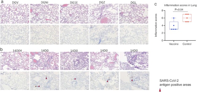
Fig. 5.
Histopathology in the lungs at day 7 post-SARS-CoV-2 challenge. H&E and immunohistochemistry to detect SARS-COV-2 antigens were performed in the vaccinated (a) and naïve (b) animals. The upper rows of (a) and (b) were H&E staining, while the lower rows of (a) and (b) were immunohistochemistry of SARS-CoV-2 detection. All images 10x (scale bar = 100 um). (c) inflammation scores in the lung were compared between the vaccinated and naïve groups. Mann–Whitney test was used for comparison. Box and whiskers with min to max were shown in the graph.

