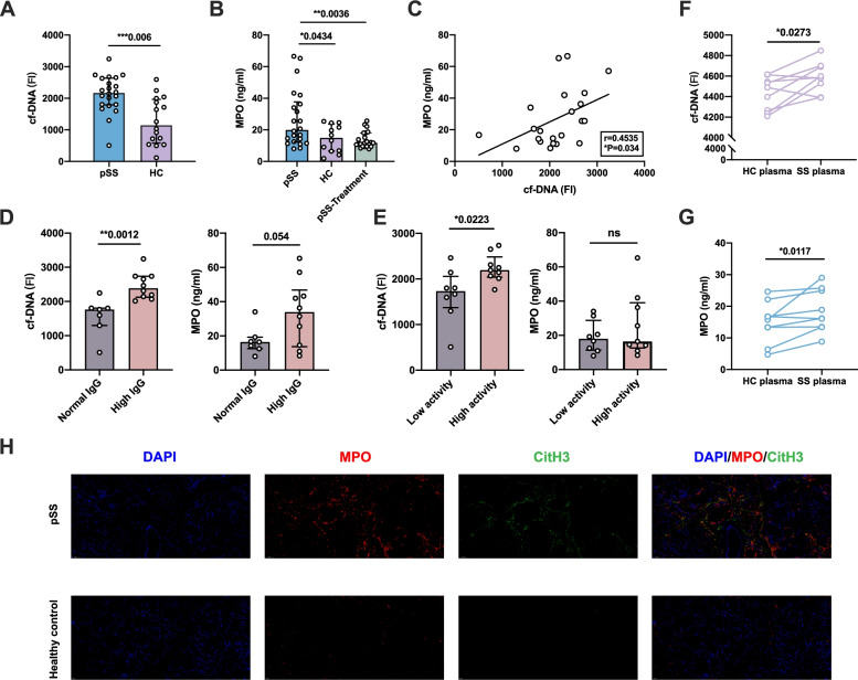
Fig. 1.
Enhanced NETosis markers in pSS patients. A Comparison of the fluorescence intensity (FI) of plasma cf-DNA between pSS patients (n=22) and matched healthy controls (n=16), each point represented the fluorescence intensity results of every subjects. B Comparison of the plasma MPO levels between pSS patients, matched healthy controls, and pSS patients after treatment, each point represented the fluorescence intensity results of every subject. C The correlation between plasma cf-DNA and MPO levels. D Comparison of plasma cf-DNA levels and MPO levels between patients with high IgG (n=10) or normal IgG (n=7) levels. E Comparison of Plasma cf-DNA levels and MPO levels between patients with high activity (n=9) or low activity (n=8), patients’ disease activity was assessed through ESSDAI (High activity, ESSDAI>5; low activity, ESSDAI≤5). F,G Comparison of plasma-stimulated neutrophils from pSS and HC to produce NETosis markers, F the fluorescence intensity of cf-DNA; G MPO production levels. H NETosis markers staining in pSS and HC labial glands (×40, blue for DAPI, red for MPO, green for CitH3). *P-value < 0.05, **P-value < 0.01, ***P-value <0.001)

