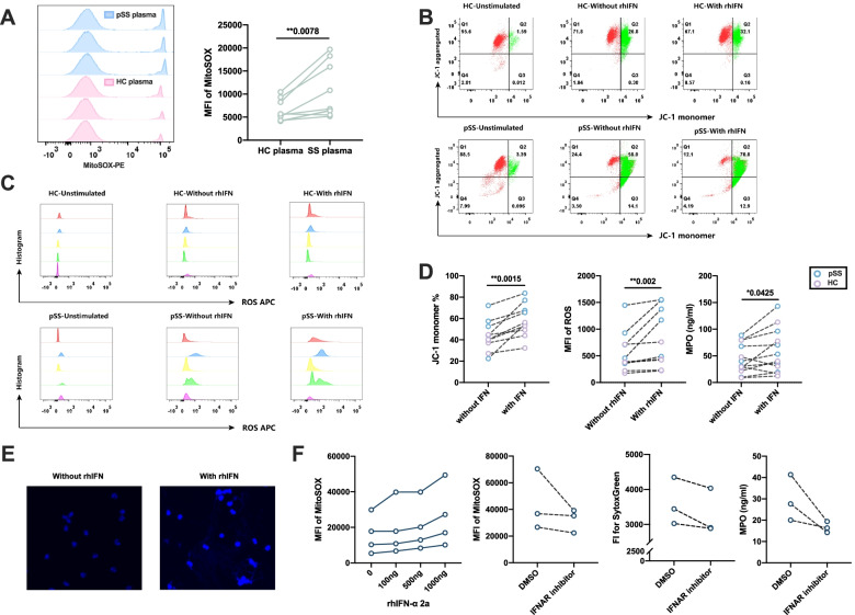
Fig. 5.
Results for rhIFN-α stimulation of neutrophils in pSS patients and healthy controls. A Results MitoSOX measurements of pSS and HC plasma stimulation of healthy neutrophils (n=9). B Representative flow cytometry results for the JC-1 staining in pSS patients and healthy controls with or without rhIFN stimulation (HC\pSS-Unstimulated represented for the freshly isolated neutrophils, pSS\HC-with or without rhIFN represented for the neutrophils stimulated with or without rhIFN). C Representative flow cytometry results of ROS production in pSS and healthy neutrophils with or without rhIFN stimulation (HC\pSS-Unstimulated represented for the freshly isolated neutrophils, pSS\HC-with or without rhIFN represented for the neutrophils stimulated with or without rhIFN, different color represented for the different subjects). D Comparison of JC-1 monomer percentage (mitochondrial damage, n=10), ROS production (n=10), and MPO concentration (n=12) between neutrophils stimulated with or without rhIFN-α. E Immunofluorescence staining of NETs-DNA (DAPI, blue) with or without rhIFN stimulation. F IFN-α receptor inhibitor reduced the mitochondrial damage (MitoSOX) and the production of NETs (SytocGreen and MPO levles). *P-value < 0.05, **P-value < 0.01, ***P-value <0.001)

