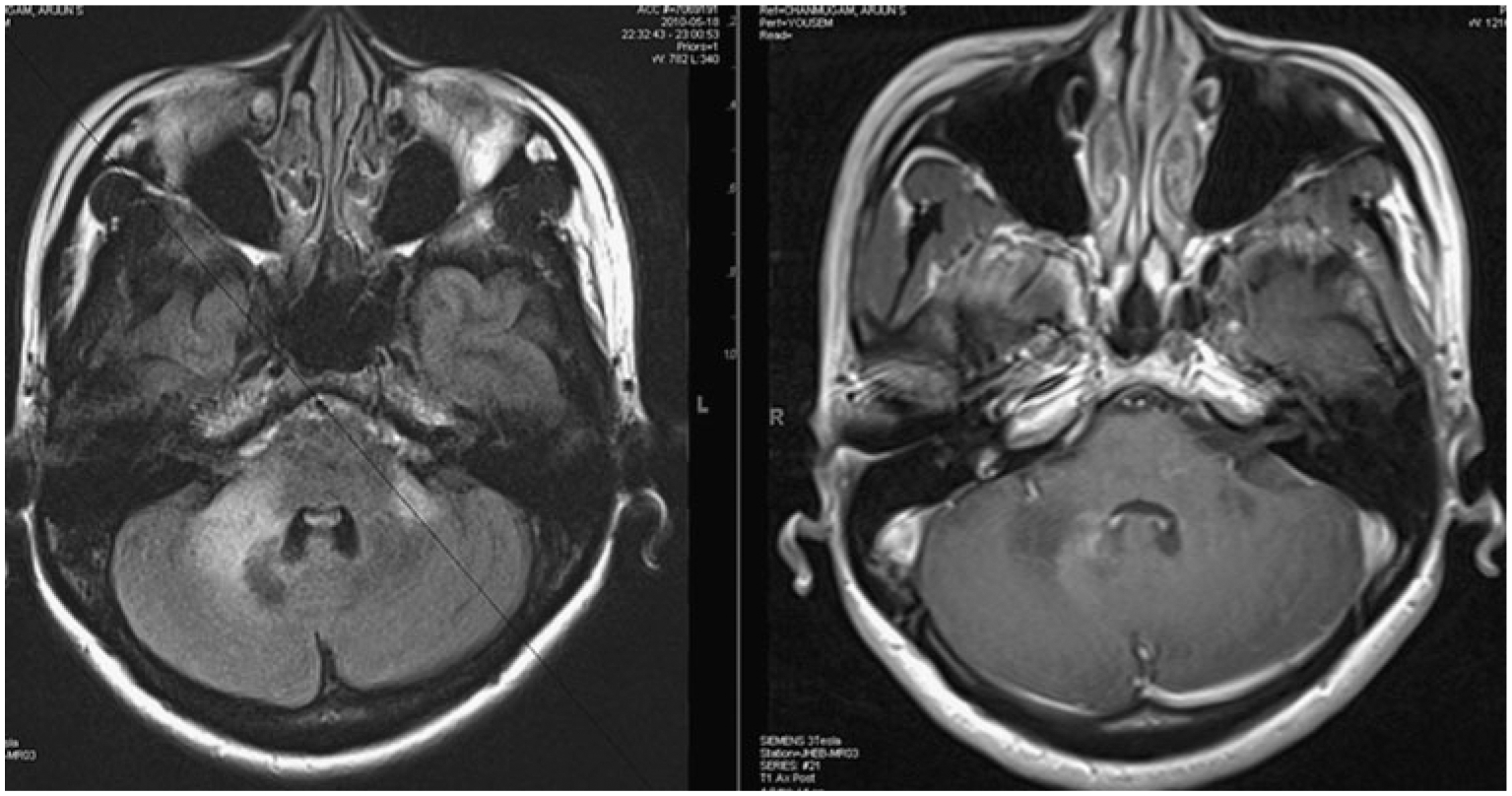
Fig. 1.

Progressive multifocal leukoencephalopathy–immune reconstitution inflammatory syndrome (PML-IRIS) with HIV infection: MRI scan with fluid-attenuated inversion recovery (FLAIR) sequence on the left shows high signal intensity lesions at the cerebellopontine junction on both sides. The corresponding contrast-enhanced T1-weighted image on the right shows an area of gadolinium enhancement in the peripheral margins of the lesion, most prominent along the medial border. This image was obtained at the time of diagnosis of PML without antiretroviral treatment and thus would be called simultaneous IRIS
