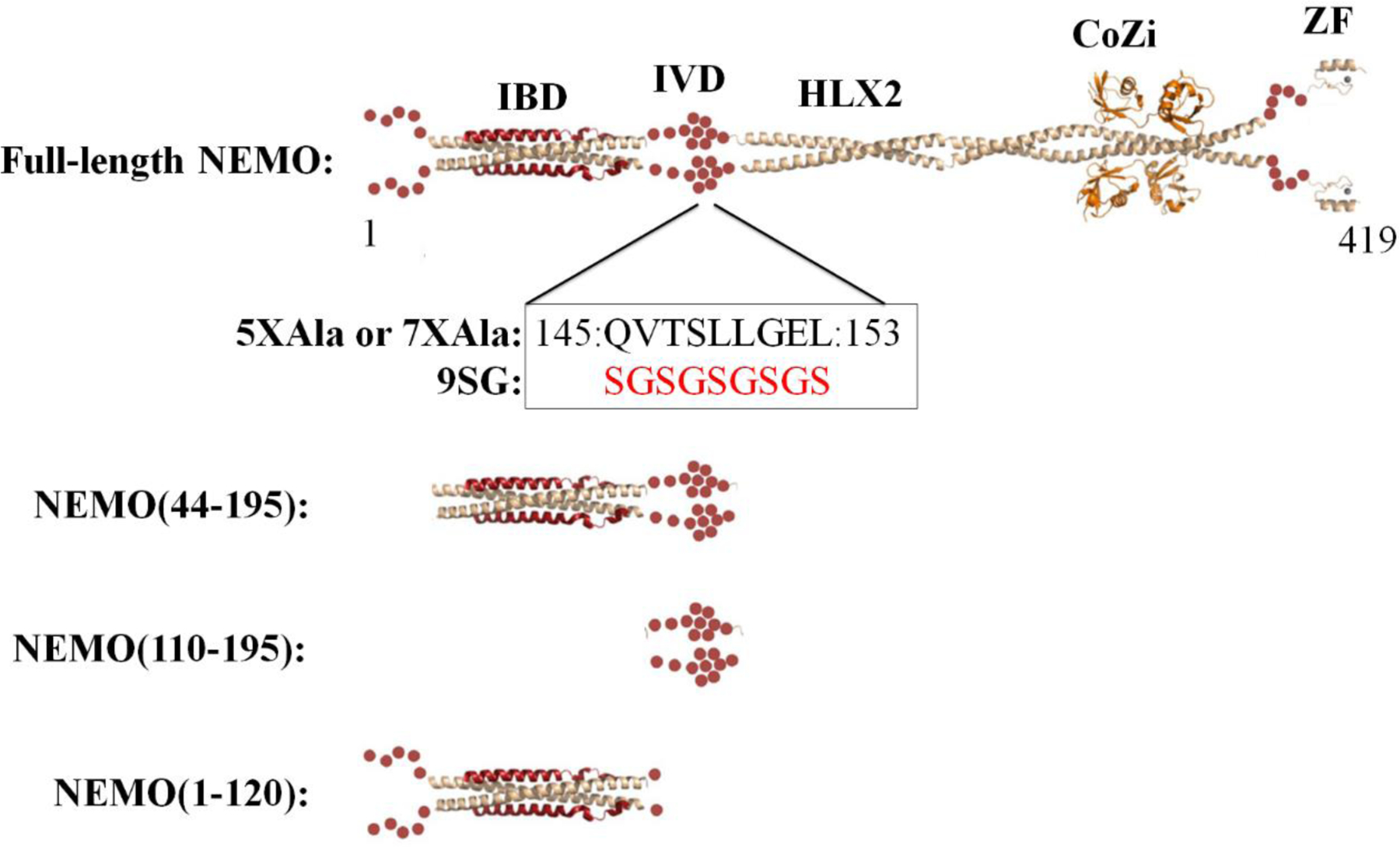
Figure 1.

Domain map of NEMO with existing experimentally determined structures of individual NEMO domains and constructs used in this study. NEMO in beige ribbon; IKKβ(701–745) in red ribbon; linear di-ubiquitin in orange ribbon; red spheres represent structurally uncharacterized regions of NEMO. IBD, IKKβ binding domain; IVD, intervening domain; HLX2, coiled-coil 2; CoZi, leucine zipper; ZF, zinc finger.
