FIG 5.
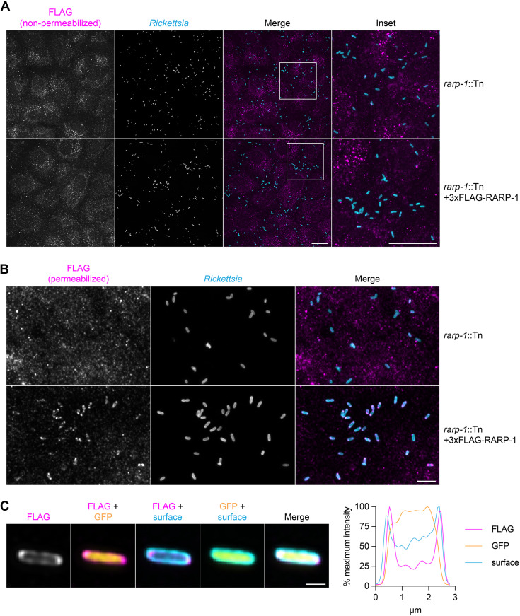
RARP-1 resides within R. parkeri. (A) Images of rarp-1::Tn (top) and rarp-1::Tn + 3×FLAG-RARP-1 (bottom) bacteria during infection of A549 cells. Samples were stained for FLAG (magenta) and the bacterial surface (cyan) without permeabilization of bacteria. Scale bars, 20 μm. (B) Images of rarp-1::Tn (top) and rarp-1::Tn + 3×FLAG-RARP-1 (bottom) bacteria during infection of A549 cells. The bacterial surface (cyan) was stained prior to permeabilization by lysozyme and detergent and subsequent staining for FLAG (magenta). Scale bar, 5 μm. (C) Subcellular localization of 3×FLAG-RARP-1 in a representative rarp-1::Tn + 3×FLAG-RARP-1 bacterium during infection of A549 cells. The bacterial surface (cyan) was stained prior to permeabilization by lysozyme and detergent and subsequent staining for FLAG (magenta). GFP (yellow) demarcates the bacterial cytoplasm. Scale bar, 1 μm. A pole-to-pole 0.26-μm-width line scan (right) was generated for FLAG, GFP, and the bacterial surface.

