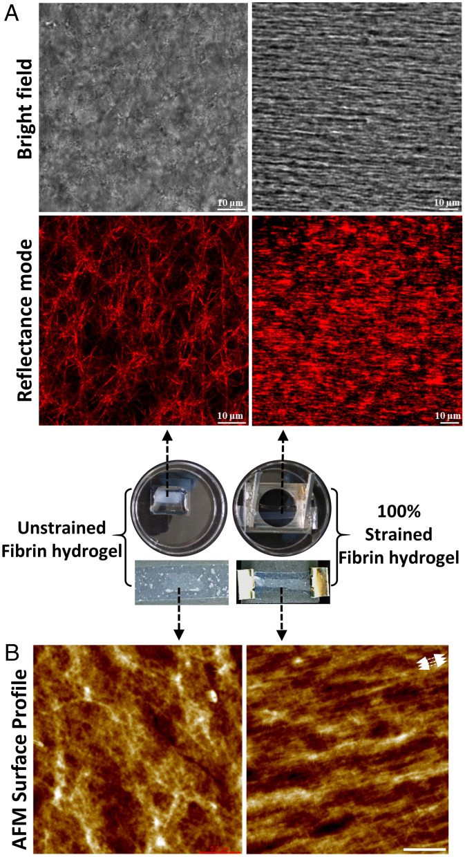
Fig. 1.
Morphology changes of fibrin under mechanical strain. (A) Transmitted light (Top) and reflectance (Bottom) confocal micrographs of unstrained (Left) and 100% strained fibrin networks (Right). Strain was applied in the horizontal laboratory frame. Images are taken ∼50 µm deep into network from the top surface, ∼1 mm away from the PDMS-fibrin interface where the strain was applied. (B) AFM topography images of unstrained and 100% strained fibrin (white arrows indicate the stretching direction), (Scale bar, 5 µm) on the top surface. The middle photos show samples corresponding to unstrained and 100% strained fibrin samples.

