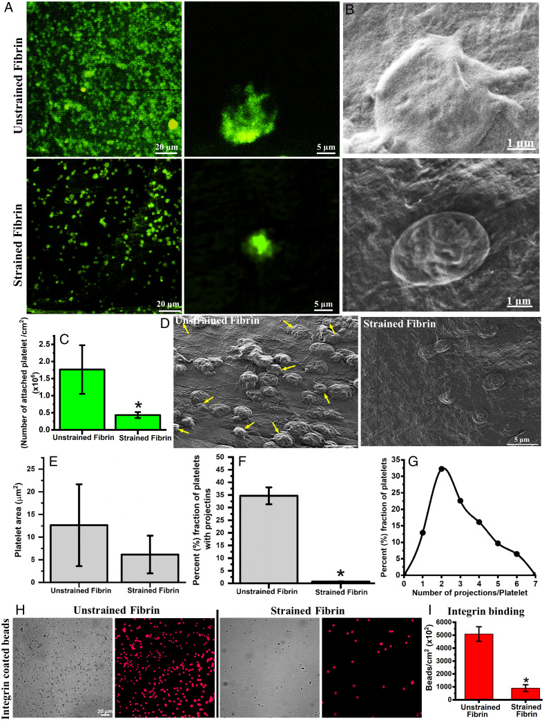
Fig. 4.
Attachment, morphology and αIIbβ3 integrin binding of platelets on different fibrin surfaces. (A) Fluorescence micrographs of attached platelets stained with Calcein on unstrained and strained fibrin gels. (B) SEM micrographs of platelets on unstrained and strained fibrin showing distinct morphology. (C) Quantification of attached platelets on unstrained and strained fibrin. (D) SEM micrographs of platelets on unstrained and strained fibrin surface (arrows indicate platelets with projections). (E) Quantification of platelet spreading (projected area) on unstrained and strained fibrin hydrogel. (F) and (G) Fraction of attached platelets with multiple projections and distribution profile of number of projections per platelets on unstrained fibrin. (H) Fluorescence micrograph of bound integrin-coated microbeads on unstrained and strained fibrin. (I) Quantitative plot of bound integrin coated microbeads on unstrained and strained fibrin. Statistically significant differences (P < 0.05) compared to unstrained fibrin is indicated by an asterisk.

