FIGURE 3.

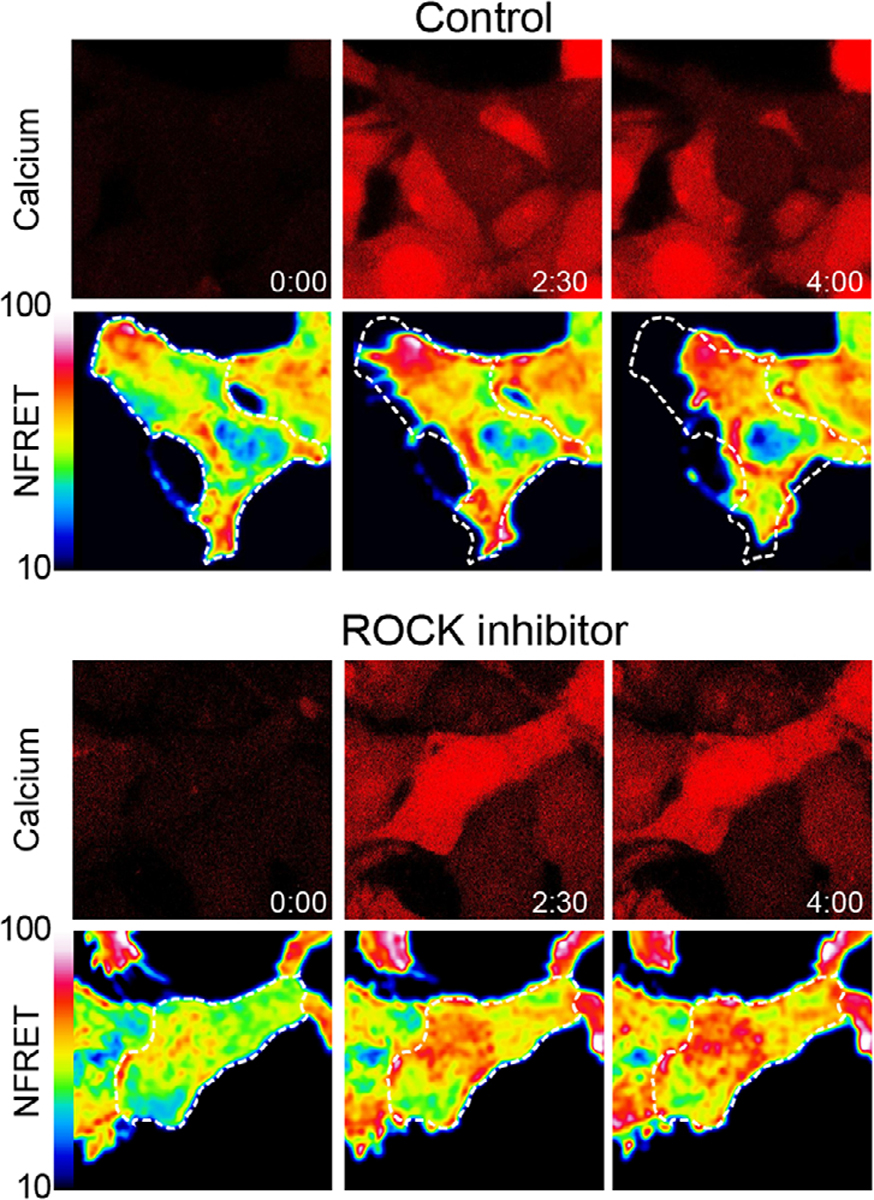
Stimulation of transient receptor potential vanilloid 4 (TRPV4) leads to RhoA activation and ROCK-dependent cytoskeletal changes. Time-lapse confocal microscopy images of stable, inducible TRPV4-expressing HEK293T cells (T-Rex-TRPV4) expressing a RhoA FRET biosensor (RhoA2G-mVenus-mTFP) and loaded with Cal590 calcium indicator demonstrate dynamics of calcium influx, RhoA activation, and cytoskeletal changes after stimulation of TRPV4. In the top panel, addition of the TRPV4 agonist GSK1016790A (100 nM) leads to calcium influx and increased RhoA activation (indicated by increased normalized FRET (NFRET)) followed by cellular contraction over 4 min. In the bottom panel, addition of the ROCK inhibitor Y-26732 (10 μM) blocks cytoskeletal changes downstream of TRPV4-mediated RhoA activation. Detailed methods were described previously[39]
