Figure 3.
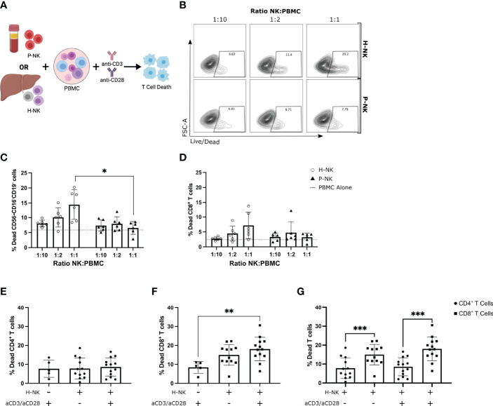
Hepatic NK cells but not peripheral blood NK cells dose-dependently kill activated allogenic peripheral blood CD8+ T cells. PBMCs cultured alone or with magnetically sorted hepatic NK cells (H-NK) or peripheral blood NK cells (P-NK) cells for 24 hr with or without anti-CD3/28 activation stimulation (n = 6-13). (A), Schematic of experimental design. (B), Representative flow plots of % dead CD56-CD16-CD19- cells at each indicated ratio for both H-NK and P-NK cocultures. (C, D), Percent dead CD56-CD16-CD19- cells (C) and CD56-CD16-CD19-CD8+ cells (D) after 24 hr co-culture with H-NK (clear circle) or P-NK (triangle). Grey dashed line represents PBMCs cultured alone. (E–G), Percent dead CD4+ T cells (E) and CD8+ T cells (F) post-PBMC culture alone or with H-NK cells and/or anti-CD3/28 activation (n = 12-13) (G), Data from (E, F) directly comparing CD4+ T cell and CD8+ T cell death post-coculture with or without anti-CD3/CD28 activation. Data analysed using a 2-Way ANOVA (C, D), a one-way ANOVA (E, F) or a paired t test (G). *p<0.05, ** p<0.01, and ***p<0.001.

