Figure 1.

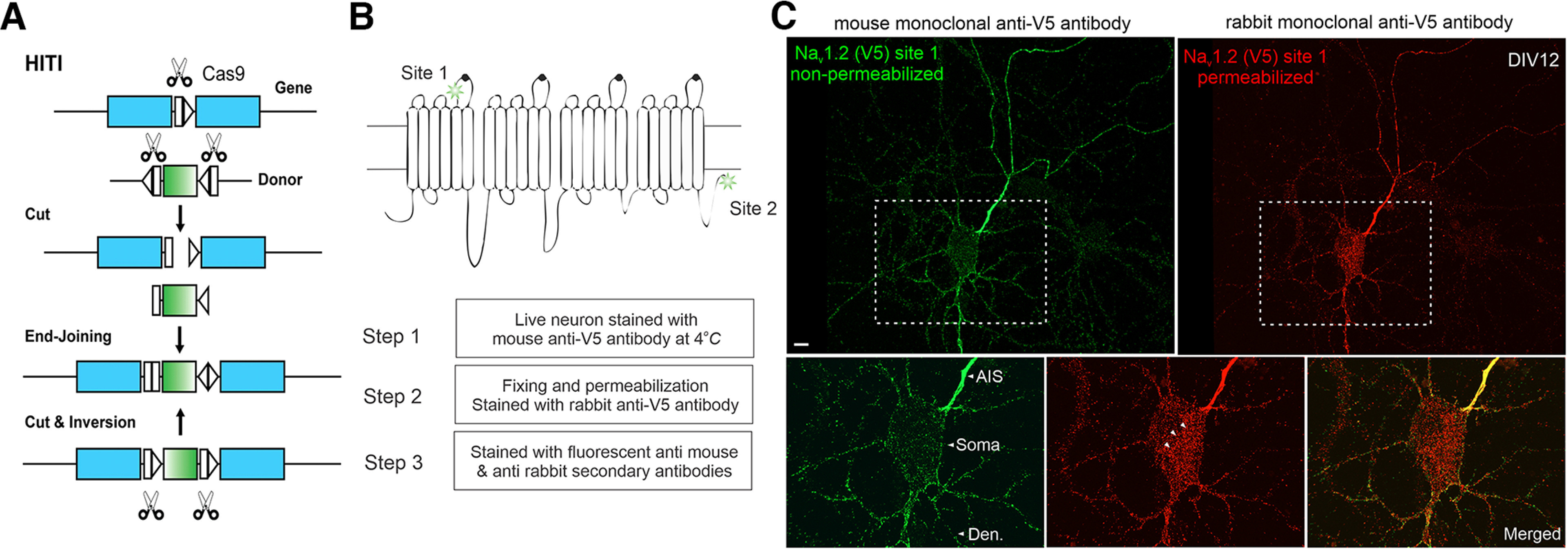
The HITI knock-in strategy and surface staining of V5-labeled Nav1.2 in cultured hippocampal neurons. A, The schematics for the HITI strategy (Suzuki et al., 2016). The donor DNA fragment has two gRNA cutting sites flanking the tag cDNA. After cutting and end-joining, if the fragment is inserted into the genome in the right direction, the two gRNA cutting sites will be inactivated. If not, the cutting and end-joining process will continue until it is inserted in the right direction. B, Top, Two gRNA targeting sites of Nav1.2 used in this study. Site 1 is at the extracellular loop between segment 5 and 6 of domain I. Site 2 is at the intracellular region near the C-terminus. Bottom, With V5 insertion at Site 1, we performed three-step immunofluorescence staining shown in C. C, Nonpermeabilized (green) and permeabilized immunofluorescence staining (red) images of Nav1.2 for the same neuron. Bottom, Zoomed-in views of the soma region (rectangle region with dashed lines). Arrowheads indicate labeled intracellular Nav1.2 vesicles (red), which were absent in the nonpermeabilized staining (green). Scale bar, 10 µm.
