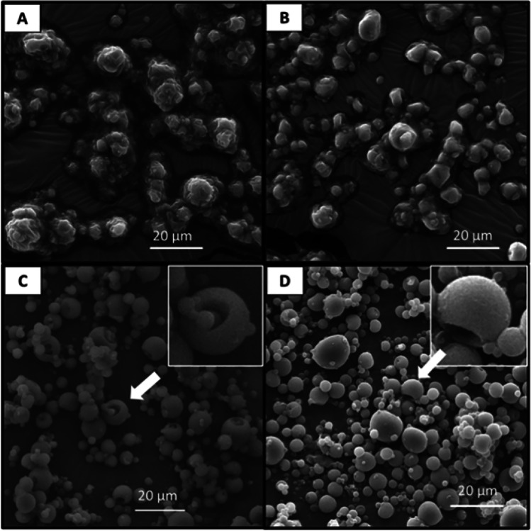
Fig. 4.
Scanning electron microscope (SEM) images showing particle morphology of the following mannitol-dextran (MD) spray dried powders: (A) MD (1:3)–40 kDa dextran, (B) MD (1:3)–500 kDa dextran (C) MD (3:1)–40 kDa dextran and (D) MD (3:1)–500 kDa dextran. Arrows indicate particle dimpling and indentation likely due to hollow shell formation. All images were captured at 2000 X magnification with a scale bar representing 20 µm in length.

