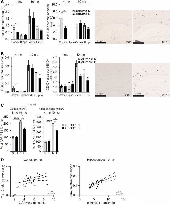
Fig. 6.
Hypoxia treatment decreases brain neuroinflammatory responses in the APP/PSEN1 mice. A IBA1-positive area total area and IBA1-positive area in relation to the Aβ deposit-affected area. Aβ-positive deposits were detected by 6E10 antibody. Example pictures of histological stainings of consecutive sections of IBA1 and 6e10 in cortex and hippocampus, respectively. B CD45-positive total area and CD45-positive area in relation to Aβ area detected by 6E10 antibody. Example pictures of histological stainings of consecutive sections of CD45 and 6E10 in cortex and hippocampus, respectively. C qPCR analysis of the mRNA levels of Trem2 in the cortex and the hippocampus of 4 and 10 mo APP/PSEN1 mice. D Correlation of the Trem2 mRNA relative expression with the Aβ amount in the cortex and hippocampus of the 10 mo APP/PSEN1 mice. Data are means ± SEM. *P < 0.05 in T-test, ##P < 0.01, ####P < 0.0001 in 2-way ANOVA. n = 14 WT N 4 mo, n = 12 WT H 4 mo, n = 8 APP/PSEN1 N 4 mo, n = 9–10 APP/PSEN1 H 4 mo, n = 14 WT N 10 mo, n = 11 WT H 10 mo, n = 8–9 APP/PSEN1 N 10 mo, n = 9–11 APP/PSEN1 H 10 mo. Hippo hippocampus, H hypoxia, mo months old, N normoxia

