Fig. 2.

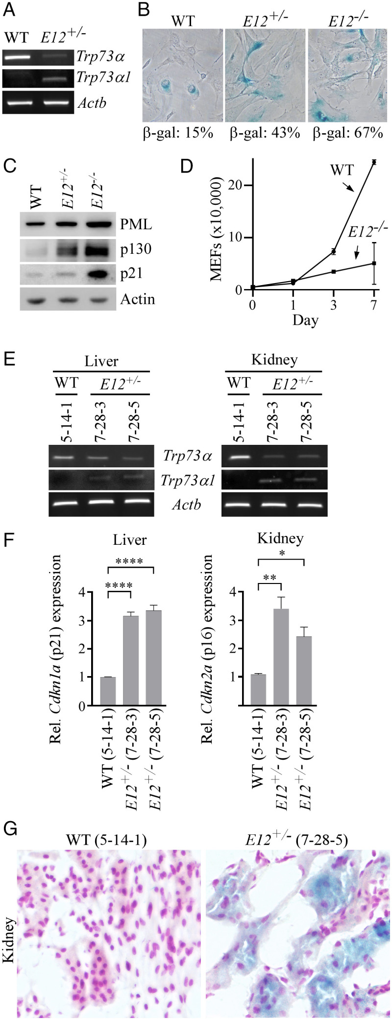
The loss of E12 in MEFs and mouse tissues leads to increased cellular senescence. (A) The level of Trp73α, Trp73α1, and Actb transcripts was measured in WT and E12+/− MEFs. (B) SA-β-galactosidase staining was performed with WT, E12+/-, and E12−/− MEFs. The percentage of SA-β-gal-positive cells was shown in each panel. (C) The level of PML, p130, p21, and actin proteins was measured in WT, E12+/-, and E12−/− MEFs. (D) Growth curves were performed with WT and E12−/− MEFs over 7 d. (E) The level of Trp73α, Trp73α1, and Actb transcripts was measured in liver and kidney tissues from age- and sex-matched WT and E12+/− mice (100 wk; Female). (F) qPCR was used to analyze relative Cdkn1a (p21) and Cdkn2a (p16) expression in liver and kidney tissues from age- and sex-matched WT and E12+/− mice (100 wk; Female). One-way ANOVA was used to calculate P values. *P < 0.05; **P < 0.01; ****P < 0.0001. (G) SA-β-galactosidase staining was performed with kidney tissues from age- and sex-matched WT and E12+/− mice (100 wk; Female).
