Figure 1.

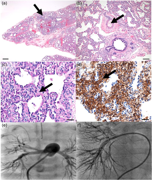
Lung biopsy at 5 weeks of life (a–d) showing: (a) Lung immaturity and moderate alveolar simplification (late saccular stage of development) with alveolar septal widening by oval mesenchymal cells. Bronchiole and alveolar duct closely approaching the pleural surface. Hematoxylin and eosin stain. Scale bar = 500 microns. (b) Pulmonary artery with moderate medial thickening and periarterial adventitial thickening. Hematoxylin and eosin stain. Scale bar = 100 microns. (c–d) Alveolar septal thickening by oval mesenchymal cells with increase in (c) cytoplasmic glycogen by Periodic acid Schiff stain, and (d) vimentin expression by immunohistochemical stain. Scale bar = 30 microns for both images. Angiography at cardiac catheterization (e, f) showing: (e) Contrast in the main pulmonary artery with markedly increased size of distal vessels and pruning with modest capillary blush at 5 weeks of life. (f) Contrast in the right pulmonary artery with relatively normal central and peripheral vessels without significant pruning and decreased capillary blush at 15 months of life, improved from prior
