
Figure 2.
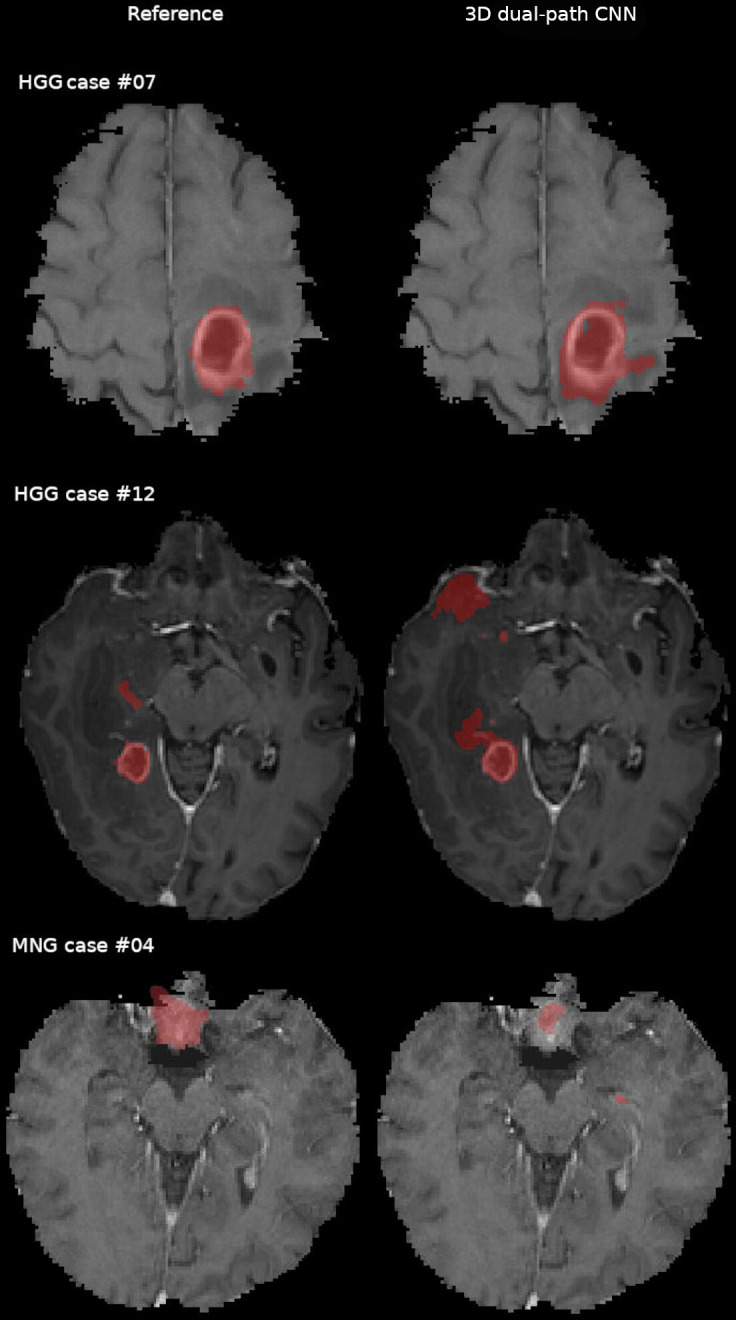
Comparison of the expert segmentation (reference) and the three-dimensional (3D) dual-path CNN tumour core segmentation in the in-house data for high-grade glioma (HGG) and meningioma (MNG) cases overlaid on contrast enhanced T1 weighted. Voxels misclassified by the 3D dual-path CNN are visible in HGG cases #07 and #12 (top and middle row). The 3D dual-path CNN failed to correctly outline the tumour and included normal brain structures in the left medial temporal lobe for MNG case #04 (bottom row). CNN, convolutional neural network.