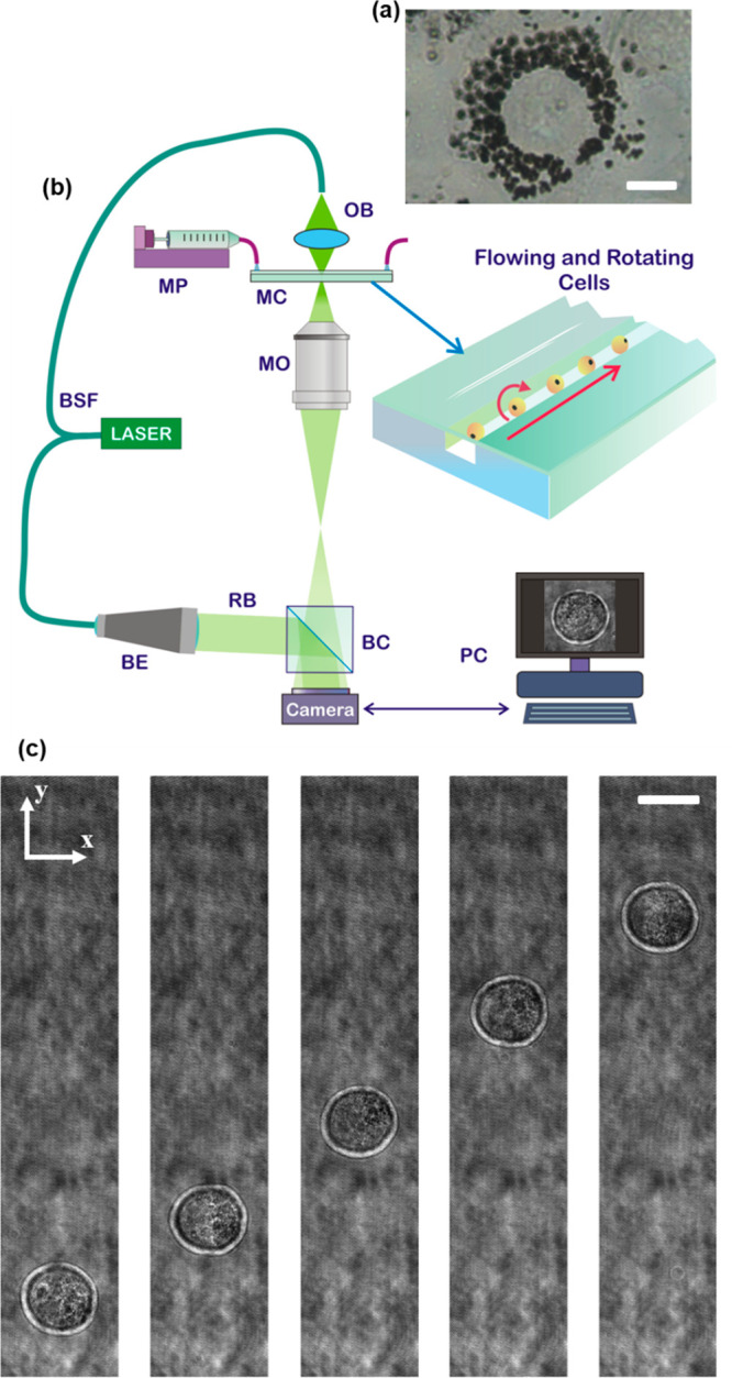
Figure 1.

(See Supporting Movie 1) Two-dimensional imaging of NIH-3T3 cells. (a) Inverted microscope image of an NIH-3T3 cell after 48 h from the nGO adding in DMEM medium. The internalized nGO (black) distributes around the nucleus. Scale bar is 10 μm. (b) Sketch of the opto-fluidic recording system based on a DH microscope in off-axis configuration. BSF, beam splitter fiber; MP, microfluidic pump; MC, microfluidic channel; OB, object beam; MO, microscope objective; BE, beam expander; RB, reference beam; BC, beam combiner; PC, personal computer. (c) Five cuts of digital holograms of the same NIH-3T3 cell at different time frames after 24 h from the nGO adding in DMEM medium. Cell flows along the y-axis and rotates around the x-axis. Scale bar is 20 μm.
