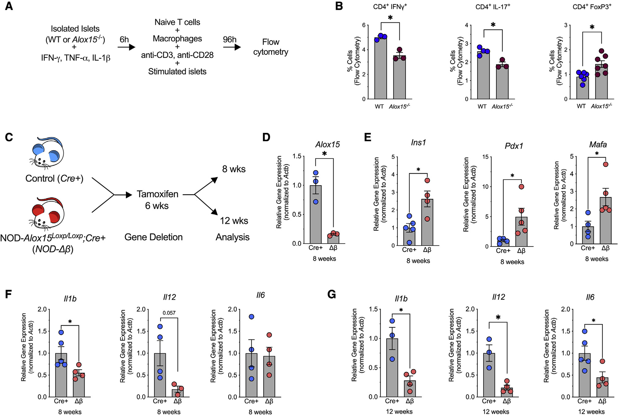
Figure 1. Alox15 deletion in the β cell enhances expression of β cell genes and reduces expression of proinflammatory genes.

(A and B) Wild-type (WT) and Alox15−/− islets were stimulated with IFN-γ, IL-1β, and TNF-α for 6 h. After stimulation, the islets were co-cultured with WT macrophages and WT naive T cells for 96 h.
(A) Experimental schematic.
(B) T cell activation was quantified by flow cytometry for CD4+IFN-γ+, CD4+IL-17+, and CD4+FoxP3+ T cells. N = 3–7 biological replicates.
(C–G) NOD-Alox15loxP/+ mice were crossed with NOD-Pdx1PB-CreER™ mice to generate NOD-Cre+ and NOD-Δβ mice. At 6 weeks of age, the mice were administered 3 daily intraperitoneal injections of 2.5 mg of tamoxifen.
(C) Experimental design.
(D–F) Quantitative RT-PCR from isolated islets at 8 weeks of age for the indicated genes.
(G) Quantitative RT-PCR from isolated islets at 12 weeks of age for indicated genes. N = 3–5 biological replicates.
Data are expressed as the mean ± SEM. *p < 0.05.
