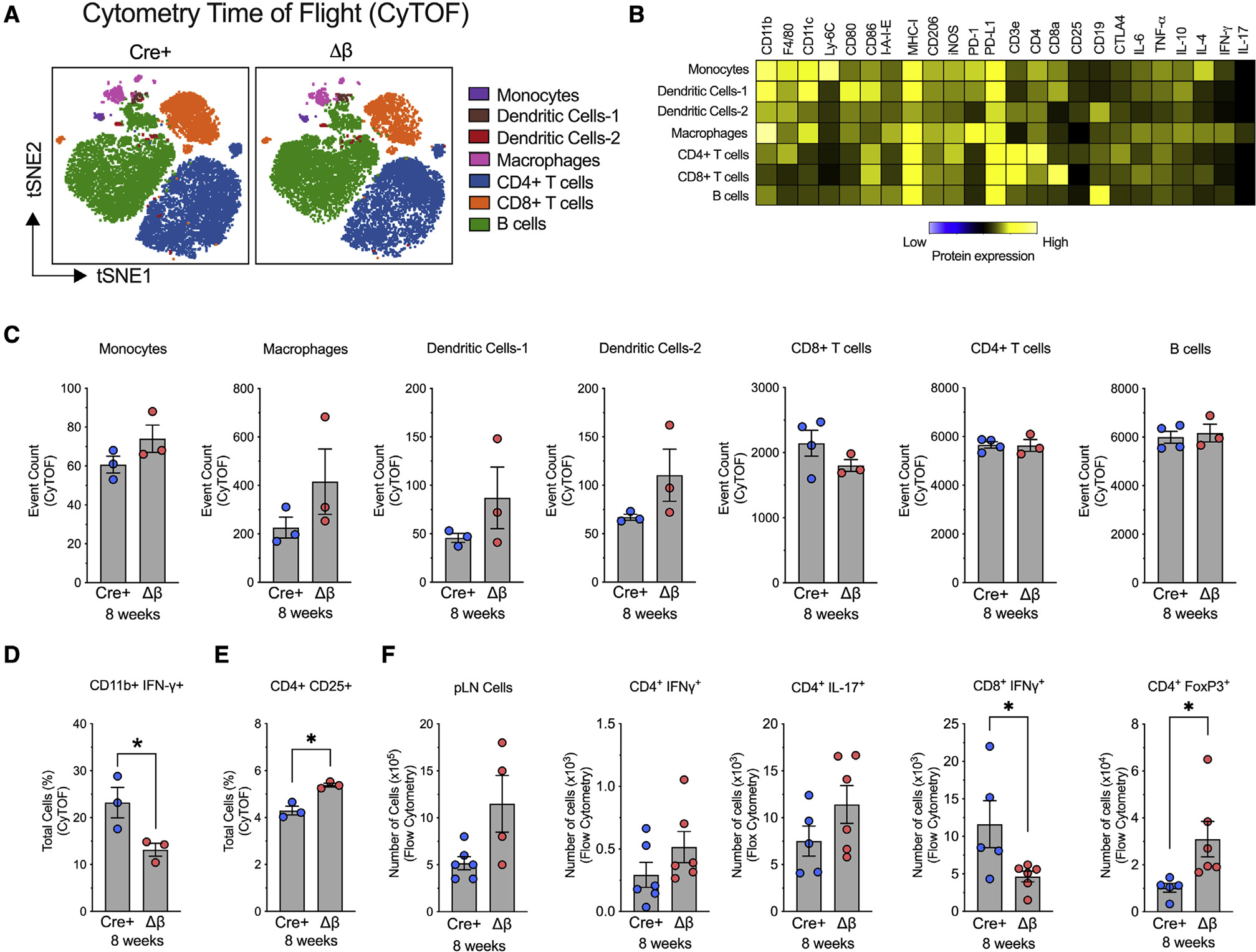
Figure 3. Alteration in immune cell subtypes following deletion of Alox15 in β cells of NOD mice.

pLNs were isolated from NOD-Cre+ and NOD-Δβ mice at 8 weeks of age, dissociated into single cells, and analyzed by cytometry by time of flight (CyTOF) or flow cytometry. N = 3–4 individual animal replicates.
(A) tSNE maps of CD45+ immune cell populations in NOD-Cre+ and NOD-Δβ pancreata by CyTOF. Cells are colored by their FlowSOM-based cluster-assignment cell.
(B) Heatmap of protein expression levels across all populations identified by FlowSOM.
(C) Quantification of each immune cell type identified by FlowSOM-based clusters.
(D) Quantification of CD11b+IFN-γ+ cells.
(E) Quantification of CD4+CD25+ cells.
(F) Flow cytometry of pLNs of CD4+ and CD8+ T cell populations.
Data are expressed as the mean ± SEM. *p < 0.05.
