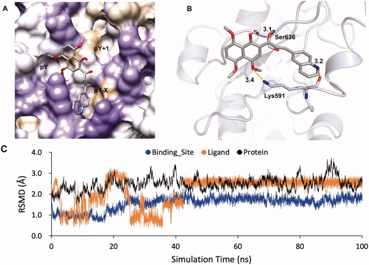
Figure 7.
Docking model of KL-6 binding to the STAT3 SH2 domain (PDB: 1BG1) generated by Autodock Vina. (A) KL-6 was coloured by atom type. The surface of SH2 domain was coloured according to hydrophobic property (the most polar residues are medium purple while the most hydrophobic are tan). (B) STAT3 protein was represented as cartoon. The key residues were shown as sticks. Hydrogen bonds were represented by yellow dashes. (C)The RMSDs of the STAT3- KL-6 complex obtained during 100 ns of MD simulation.

