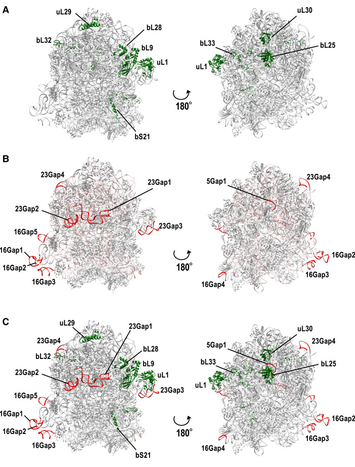
FIGURE 6.
Missing rRNA regions and missing ribosomal proteins in CPR bacteria map to the surface of the 3D E coli ribosomal structure. A three-dimensional structural model of the E. coli K-12 ribosome (PDB ID: 5U9G) was used for the analysis. (A) Ribosomal proteins missing in some or all CPR bacteria (see Fig. 2; Supplemental Fig. S3) are shown in green. (B) 16S, 23S, and 5S rRNA regions missing in CPR bacteria (see Fig. 5) are colored red. Entire rRNA regions are colored light pink. (C) Mixed view of A and B.

