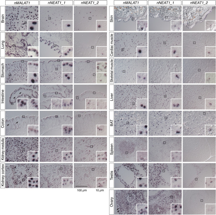
FIGURE 3.
Expression of nMALAT1 and nNEAT1 in various tissues of naked mole-rats. Expression patterns of nMALAT1, nNEAT1_1, and nNEAT1_2 in brain, lung, stomach, intestine, colon, kidney, skin, cardiac muscle, skeletal muscle, liver, brown adipose tissue (BAT), spleen, testis, and ovary of naked mole-rats detected by in situ hybridization. All tissues are from a nonbreeder male animal (1 yr 8 mo) except for the ovary, which is derived from a non-queen female animal (1 yr 11 mo). Black squares represent areas shown in insets at a higher magnification. Arrows in the nMALAT1 panels indicate the cells that do not express nMALAT1. Scale bars are 100 µm for large panels and 10 µm for insets.

