Figure 5.
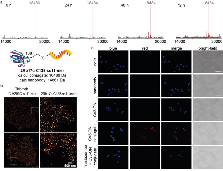
Bioconjugate integrity studies and cell imaging experiments. a) LC‐MS based conjugate integrity study of the anti‐HER2 nanobody conjugate 2Rb17c‐C138‐ss11‐mer in the presence of 10 % human plasma with deconvoluted mass spectra of samples taken at specified time points. No detectable degradation of the DNA‐nanobody conjugate was observed and an intact 2Rb17c‐C138‐ss11‐mer conjugate was identified by LC‐MS after 72 h. For full LC‐MS spectra see Figures S60–S63. b) Super‐resolution images of HER2 receptors on SKBR3 breast cancer cells obtained by DNA‐PAINT method. 2Rb17c‐C138‐ss11‐mer and Thiomab LC‐V205C‐ss11‐mer were used as probes. Scale bars represent 1000 nm (top images) or 500 nm (bottom images). c) Epifluorescence microscopy imaging of the HER2 receptor‐mediated internalization of the 2Rb17c‐C138‐Cy3‐3′‐ss25‐mer conjugate on SKBR3 breast cancer cells. Obtained images of SKBR3 cells incubated for 2 h: without any probe, with 2Rb17c‐C138 antibody, 5′‐NH2‐Cy3‐3′‐ss25‐mer ON, 2Rb17c‐C138‐Cy3‐3′‐ss25‐mer or with 2Rb17c‐C138‐Cy3‐3′‐ss25‐mer after 1 h incubation with Trastuzumab. Images were processed using ImageJ software, scale bars represent 100 mm. Experiments were performed two independent times. Representative data from one experiment is shown. For full experimental details see SI.

