. 2022 Jun 30;9(2):323–334. doi: 10.5455/javar.2022.i599

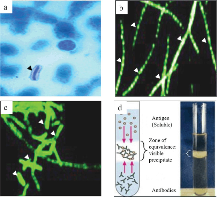
Figure 2. Staining and immunological techniques are used for the diagnosis of anthrax. (a) M’Fadyean staining, (b) FAT using monoclonal antibodies for cell wall, (c) and capsule were useful for the detection and identification of anthrax bacilli. In advanced decomposition, detection of the bacteria in the carcass is almost impossible where a precipitation test (Ascoli’s thermo precipitation test) is suitable for diagnosing anthrax (d). Black and white arrowheads indicate the bacteria after staining. Figures a–d were taken from or kindly provided by Anne Boyer (Anthrax Toxin Team Leader, CDC), De et al. [69], and Microbe Notes [83], respectively.

Figure 2.