Fig. 1.

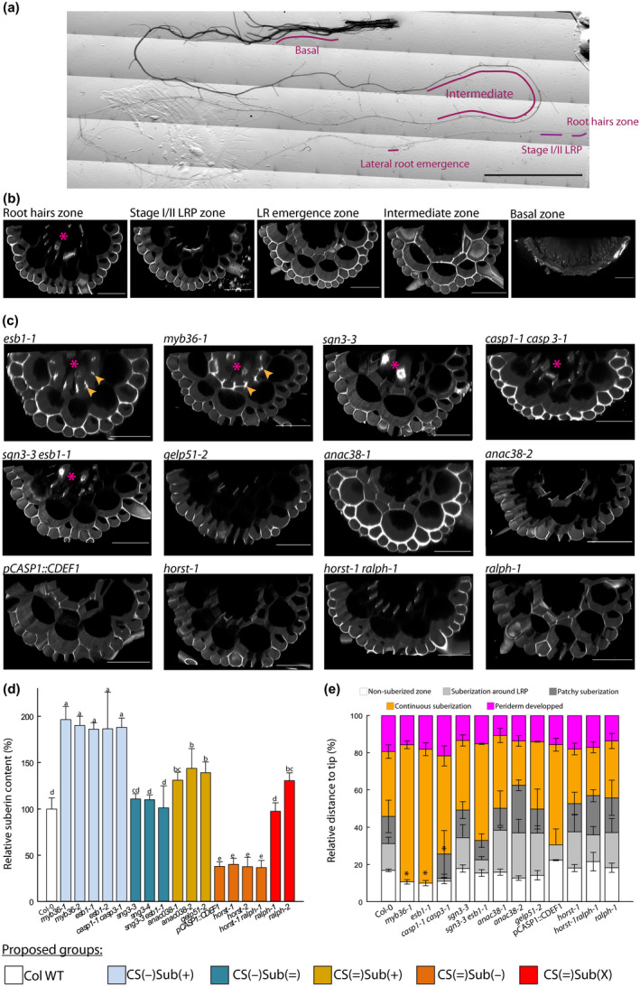
Characterization of Casparian strip permeability and suberin development. (a) Reconstituted picture of a 21 d‐old primary root, and the zones that were monitored for propidium iodide (PI) permeability. Bar, 1 cm. (b) Confocal cross‐sections of a 21 d‐old plants, PI stained, Arabidopsis root from Col‐0 at various zones: root hairs, stages I and II lateral root primordium (LRP), first lateral root (LR) emergence, intermediate, and basal. Bars, 50 µm. (c) Confocal cross‐section of PI staining in roots of 21 d‐old plant at stages I and II LRP development. Arrows highlight the staining related to ectopic deposition of cell wall polymers at the cell corners, stars indicate when the vessels are stained and hence, PI was able to penetrate through the stele. Bars, 50 µm. (d) Relative suberin content related to wild‐type Arabidopsis plants (Col‐0) of 17 Casparian strips (CS) and/or suberin mutants of 21 d‐old plants. Suberin was analyzed using gas chromatography after release by transesterification using boron trifluoride in methanol from solvent extracted root cell walls. Bars represent mean values in μg per mg dry weight ± SE (n = 3–5). *Suberin content taken from literature esb1‐2 (Baxter et al., 2009), pCASP1::CDEF1 (Barberon et al., 2016), horst‐1, horst‐2 (Hofer et al., 2008), ralph‐1, ralph‐2 (Compagnon et al., 2009). (e) Scoring of the suberin stages along the root, as a relative position from the tip ± SE, after staining with the lignin/suberin dye Auramine‐O (n = 3–5). Method detailed in Supporting Information Fig. S2. Asterisks indicate significant difference (P < 0.05) to Col‐0 plants. Colors patterns of (c) allow to visually identify the groups that are defined in the first section of the results. They are reproduced similarly over all the figures.
