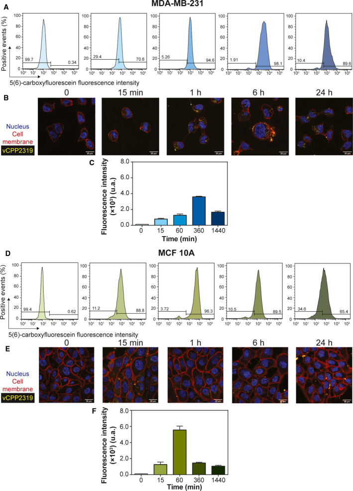
Fig. 4.

vCPP2319 internalization in human breast cells. Flow cytometry and confocal microscopy were used to evaluate the internalization of the 5(6)‐carboxyfluorescein‐labelled vCPP2319 (yellow) in MDA‐MB‐231 (A and B) and MCF 10A (D and E) cells. Cell nucleus (blue) was stained with Hoechst 33342, and cell membranes (red) were stained with CellMask™ Deep Red. The fluorescence intensity of the 5(6)‐carboxyfluorescein‐labeled vCPP2319 is also represented as function of time for treated MDA‐MB‐231 and MCF 10A cells (C and F, respectively). Error bars in C and F refer to the standard deviation obtained from three different experiments (n = 3), performed in different days with independently grown cell cultures. Scale bar represents 20 μm.
