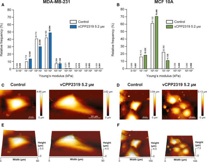
Fig. 5.

Effect of vCPP2319 on cell biomechanics assessed through AFM. Cell cultures from both cell lines were treated with 5.2 μM of vCPP2319, and live cells were analyzed in terms of their biomechanical properties using QITM mode to obtain force curves from 10 × 10 μm areas scanned over the nucleus. Young’s modulus values were obtained from force curves using the Hertz/Sneddon fit. The distribution of the Young’s modulus is displayed for MDA‐MB‐231 (A) and MCF 10A (B) cells untreated (n = 3 and n = 4, respectively) and treated with vCPP2319 (n = 3 and n = 2, respectively). The Mann–Whitney test was applied to the comparison between distributions obtained for treated and untreated samples from each cell line, and the level of significance was ****(P‐value < 0.0001) in each case. Live imaging of whole cells (100 × 100 μm) was performed also using QITM mode for untreated and treated MDA‐MB‐231 and MCF 10A cells. Representative height images (C and D) and 3D projections (E and F) for untreated and treated cells are exhibited for MDA‐MB‐231 and MCF 10A cells. Scale bar represents 10 μm in C–Control; and 20 μm in the C–vCPP2319 5.2 μM–and D.
