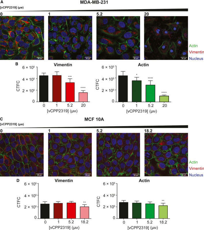
Fig. 6.

Impact of vCPP2319 on MDA‐MB‐231 and MCF 10A cytoskeleton studied by confocal microscopy. Vimentin filaments (red) and actin fibers (green) were stained with Vimentin Monoclonal Antibody (V9) eFluor 660 and CellMask™ Green Actin Tracking Stain, respectively. Cell nucleus (blue) were stained with Hoechst 33342. Images were obtained for both cells lines with increasing peptide concentrations (A and C), and corrected total cell fluorescence (CTCF) intensity was calculated for vimentin and actin in MDA‐MB‐231 (B) and MCF 10A (D) cells (n = 3). Error bars refer to the standard deviation. One‐way ANOVA followed by Tukey’s multiple comparison test was applied to infer on data significance. *P‐value ≤ 0.05; **P‐value ≤ 0.01; ***P‐value ≤ 0.001; ****P‐value ≤ 0.0001. Scale bar represents 20 μm.
