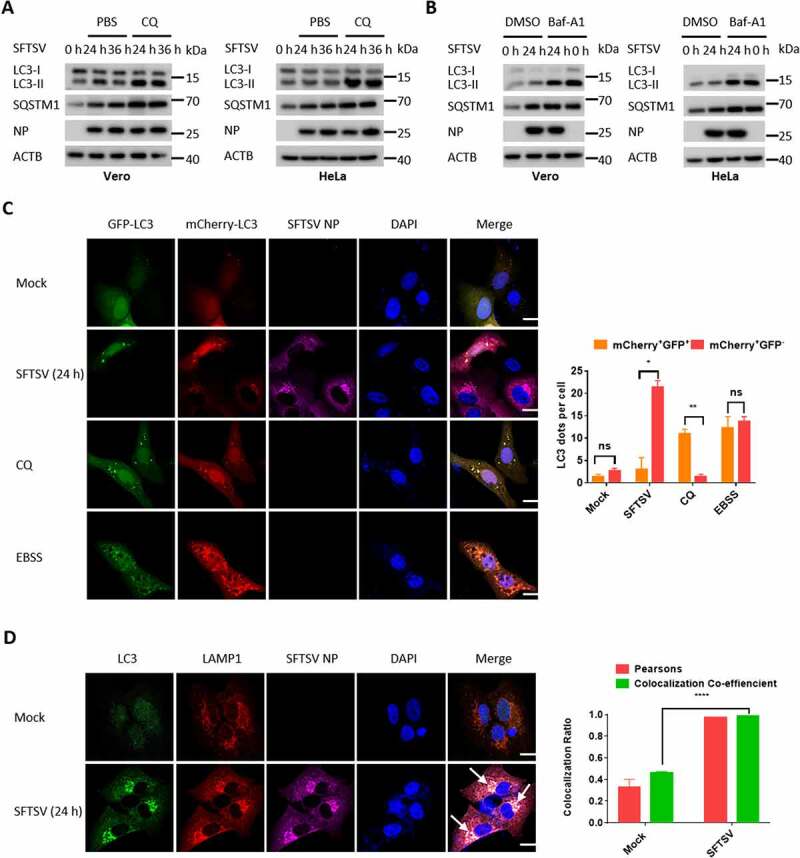
Figure 2.

SFTSV-induced autophagy is a complete process. (A) Vero or HeLa cells were mock infected or infected with SFTSV at MOI of 5 for 24 h and 36 h and then treated with CQ for 6 h. Cell lysates were evaluated via WB. (B) Vero or HeLa cells were mock infected or infected with SFTSV for 24 h and then treated with Baf-A1 for 6 h. Cell lysates were evaluated via WB. (C) Vero cells were transfected with mCherry-GFP-LC3 for 12 h and then were mock infected or infected with SFTSV, or treated with CQ (100 µM), or starved in EBSS medium for 6 h. The number of mcherry-GFP-LC3 dots in each cell was counted, and at least 20 cells were included for each group. Single plane type of images was present. Scale bar: 20 μm. (D) Vero cells were infected with SFTSV for 24 h for analyzing the colocalization of endogenous LC3 and LAMP1. Single plane type of images was present. Scale bar: 20 μm.
