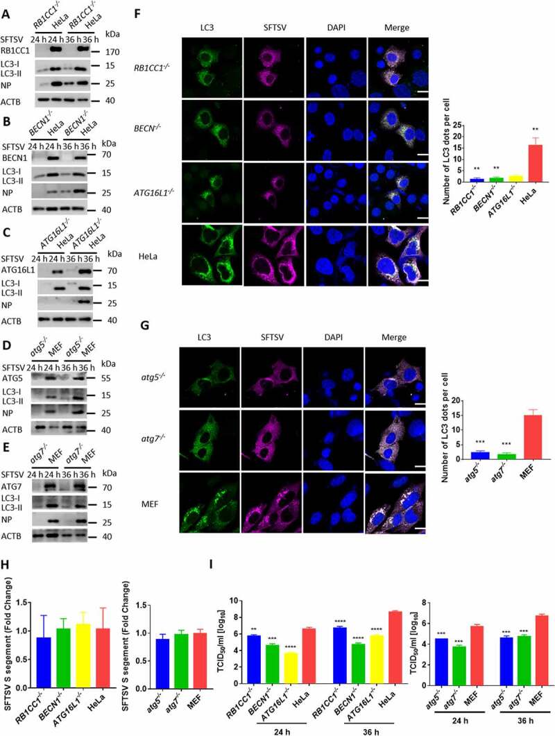
Figure 3.

Autophagy is essential for the replication of SFTSV. (A-E) BECN1, atg5, atg7, RB1CC1, ATG16L1 knockout MEF or HeLa cells and WT cells were infected with SFTSV at an MOI of 1 for 24 h and 36 h. Cell lysates were evaluated via WB. (F-G) BECN1, atg5, atg7, RB1CC1, ATG16L1 knockout MEF or HeLa cells and WT cells were infected with SFTSV at an MOI of 1 for 24 h. Nuclear DNA, endogenous LC3, and SFTSV NP were stained as blue, green, and violet respectively. Single plane type of images was present. Scale bar: 20 μm. (H) BECN1, atg5, atg7, RB1CC1, ATG16L1 knockout MEF or HeLa cells and WT cells were infected with SFTSV at an MOI of 1 for 2 h. Then cells were washed once by PBS for three times and internalized SFTSV were detected via RT-qPCR. (I) BECN1, atg5, atg7, RB1CC1, ATG16L1 knockout MEF or HeLa cells and WT cells were infected with SFTSV at an MOI of 1 for 24 h. Endpoint 10-fold dilutions of an SFTSV stock were titrated. Values presented in the graph are calculated and expressed as the log10 of TCID50 units per ml of supernatant. Error bars, mean ± SD of three experiments. Student’s t test; *p < 0.05; **p < 0.01; ***p < 0.005; ****p < 0.001.
