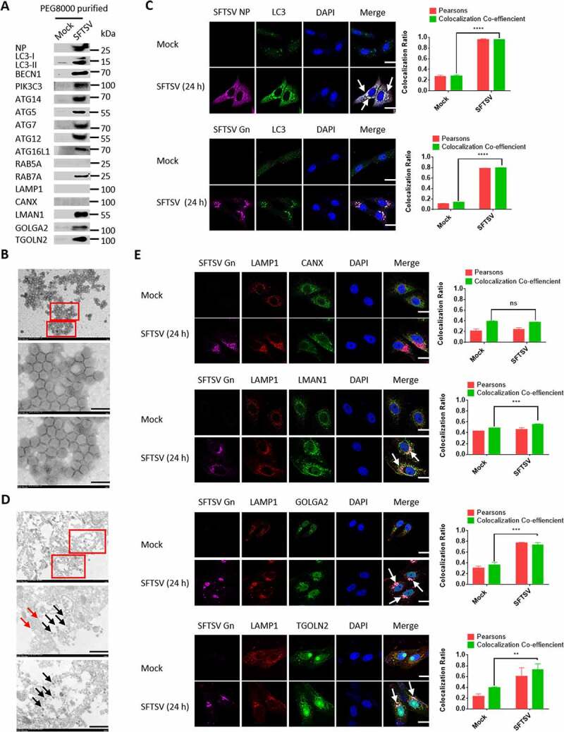
Figure 5.

The autophagosome serves as SFTSV assembly platform. (A) HeLa cells were infected with SFTSV at an MOI of 1 for 5 days and SFTSV in supernatant were harvested and purified for WB detection. (B) PEG8000 purified SFTSV particles were analyzed via electron transmission microscopy. Scale bar: 200 nm. (C) Representative images of Vero cells infected with SFTSV at an MOI of 5 for 24 h and stained for endogenous LC3, SFTSV NP, and Gn. Nuclei were stained with DAPI. Single plane type of images was present. Scale bar: 20 μm. (D) 10-nm gold particles were used to label endogenous LC3. The red arrows refer to LC3, and the black arrows refer to SFTSV virions. Single plane type of images was present. Scale bar: 500 nm. (E) Vero cells were infected with SFTSV at an MOI of 1 for 24 h. The colocalization of Gn and LAMP1 to the ER, ERGIC and Golgi was analyzed. Single plane type of images was present. Scale bar: 20 μm.
