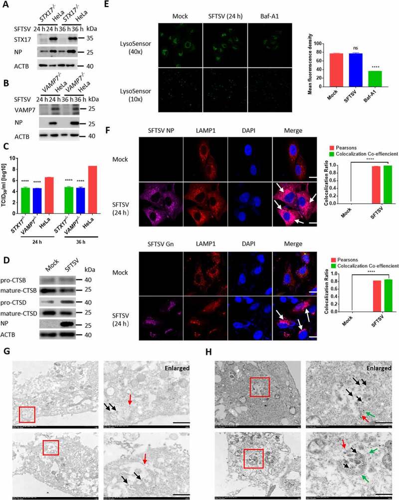
Figure 6.

Autophagic vacuoles is exploited by SFTSV for exocytosis. (A and B) STX17 orVAMP7 knockout HeLa cells and WT cells were infected with SFTSV at an MOI of 1 for 24 h and 36 h. Cell lysates were evaluated via WB. (C) STX17 or VAMP7 knockout HeLa cells and WT cells were infected with SFTSV at an MOI of 1 for 24 h and 36 h. Viral titer was measured by TCID50. (D) Vero cells were mock infected or infected with SFTSV at MOI of 5 for 24 h. Pro-form and mature-form of CTSB and CTSD were evaluated via WB. (E) Vero cells were mock infected or infected with SFTSV at MOI of 5 for 24 h or treated with Baf-A1 for 6 h. Then autolysosome acidification was determined by LysoSensor Green DND-189 staining. Single plane type of images was present. The mean density was calculated via ImageJ. Error bars, mean ± SD of three experiments. Student’s t test; *p < 0.05; **p < 0.01; ***p < 0.005; ****p < 0.001. (F) Representative images of Vero cells infected with SFTSV at an MOI of 5 for 24 h and then underwent immunofluorescence assay to mark endogenous LAMP1, SFTSV NP, and Gn. Nuclei were stained with DAPI. Single plane type of images was present. Scale bar: 20 μm. (G) 10-nm gold particles were used to label endogenous LAMP1 in SFTSV infected Vero cells. The red arrows refer to LAMP1, and the black arrows refer to SFTSV virions. Scale bar: 500 nm. (H) 10-nm gold particles were used to label endogenous LC3 and 4-nm gold particles were used to label endogenous LAMP1 in SFTSV infected HeLa cells. The red arrows refer to LC3, the green arrows refer to LAMP1 and the black arrows refer to SFTSV virions. Scale bar: 500 nm.
