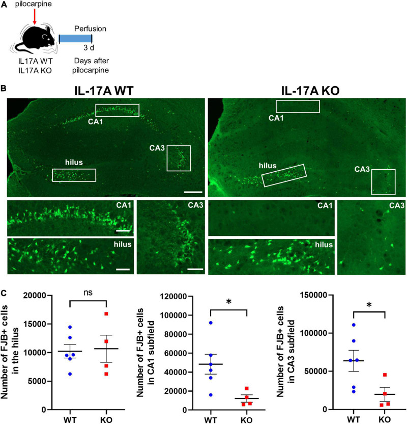
FIGURE 3.
IL-17A deficiency in mice promotes neuroprotection against pilocarpine-induced status epilepticus (SE). (A) Timeline showing the experimental design. (B) Representative microscope images showing degenerating neurons stained with Fluoro-Jade B (FJB). Magnified image areas in the hilus, CA1, and CA3 subfields are indicated as white rectangles. Scale bar = 200 μm for low magnified images, 50 μm for higher magnified images. (C) Graphs showing the number of FJB-positive cells in the hilus, CA1, and CA3 subfields of the hippocampus. Fewer degenerating neurons were observed in CA1 and CA3 subfields of the hippocampus in IL-17A KO mice compared with the WT group. n = 6 (WT) and n = 4 (KO). Detailed statistics are as follows. Hilus: Mann–Whitney U-test, p = 0.971, U = 11,500; CA1: Mann–Whitney U-test, p = 0.019, U = 1,000; CA3: Mann–Whitney U-test, p = 0.038, U = 2,000. Data are presented as mean ± SEM. *p < 0.05, NS, not significant.

