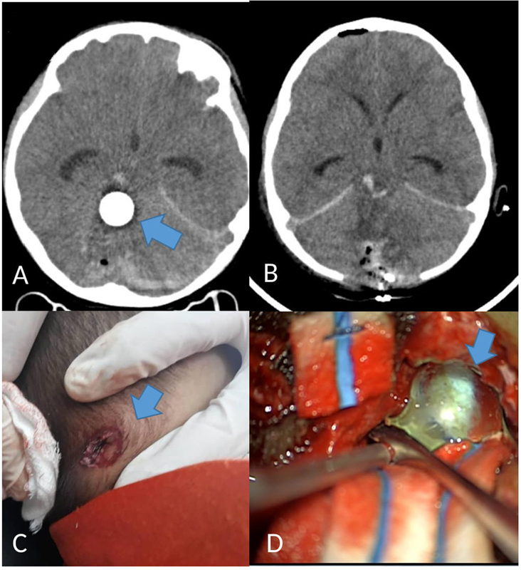
Fig. 1.

( A ) Brain CT showed a round hyperdense foreign body ( blue arrow ) at the right cerebellum, close to the mid brain. A 3-cm long tract with blood and air within was seen along its entry point. ( B ) Postoperation brain CT: less affected fourth ventricle; brain lax; Slyvian fissure opened. ( C ) Entry point of penetrating brain injury: a deep circular wound over the right occipital scalp ( blue arrow ). ( D ) Marble found and removed using tumor holding forceps ( blue arrow ).
