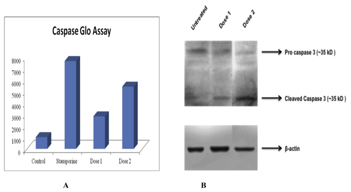
Fig. 4.
Caspase glo assay and western blotting. (A) Caspase glo assay revealed higher luminescence i.e. 5438.25 in compound six treated group at the dose of 80 μg/ml, as compare to control group which was 1021.25. The high luminescence indicates higher caspase activity and more apoptotic cells than control group. Dose 1 is 20 μg/ml and dose 2 is 80 μg/ml. (B) Western blot analysis showed expression of cleaved caspase at both the dose of ethyl iso-allocholate. The β-actin was used as loading control that expressed well in all groups nullifying the loading errors.

