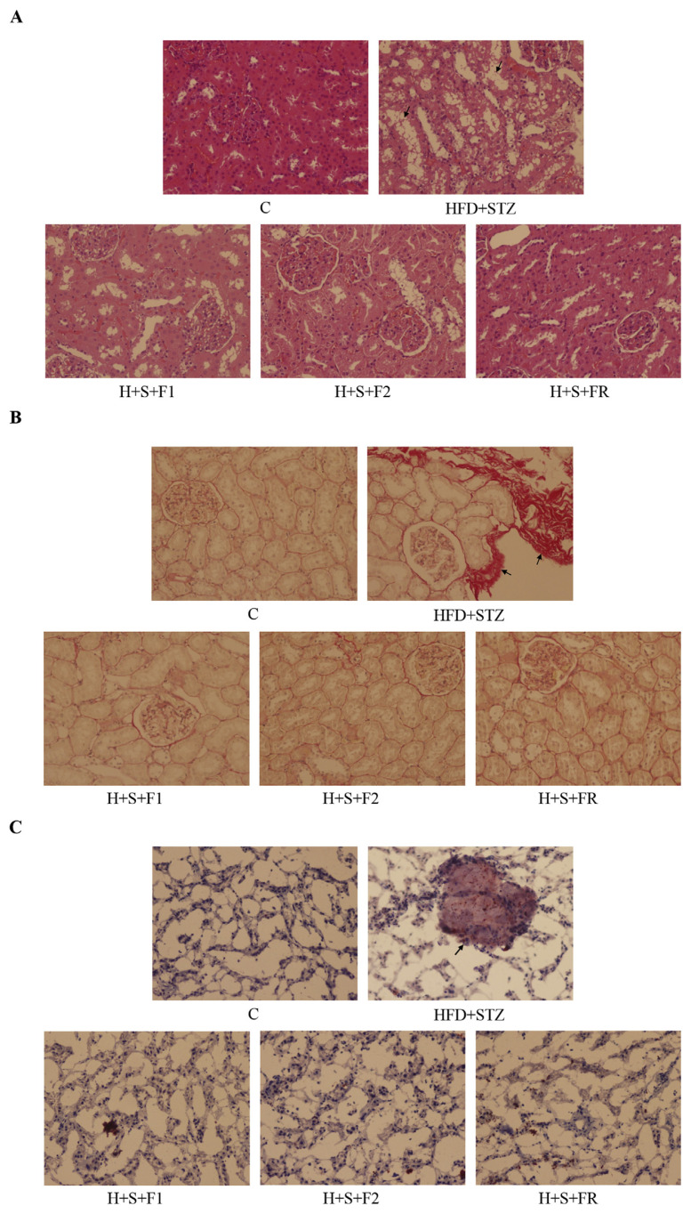
Fig. 3.
Effect of AE on the histological change. Kidneys of the control, type 2 diabetes and 0.45 mg/kg of AE subfraction-treated diabetic rats were collected, fixed, sectioned, and stained for microscopic observation (200X). (A) H–E stain (B) Sirius red stain, collagen accumulation in the red area (C) Oil red O stain, fat deposits in the red-stained area. Arrows indicate the change in diabetic kidneys.

