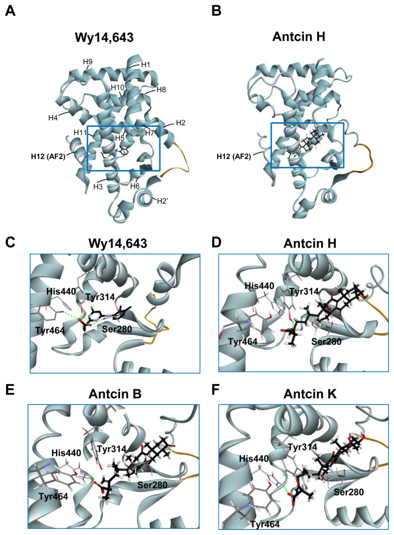
Fig. 4.
Three-dimensional structures of PPARα-LBD stabilized by the ligands. (A, B) Overall structure of Wy-14,643 (A) and antcin H (B) bound with PPARα in ribbon representation. The bound ligands are shown in stick representation. The H12 (AF2) helix is labeled. Missing residues in the H2–H2′ loop are depicted as an orange curve. (C–F) Close-up view of the Wy-14,643 (C), antcin H (D), antcin B (E), and antcin K (F) at the binding site of PPARα showing the interacting residues. Ligands are shown as black sticks, and receptor residues are shown as grey sticks. The bound ligands and receptor residues are shown in stick representation with oxygen, nitrogen, chloride, sulfur and hydrogen atoms depicted in red, blue, green, yellow, and white, respectively. Hydrogen bonds are shown as dotted green lines.

