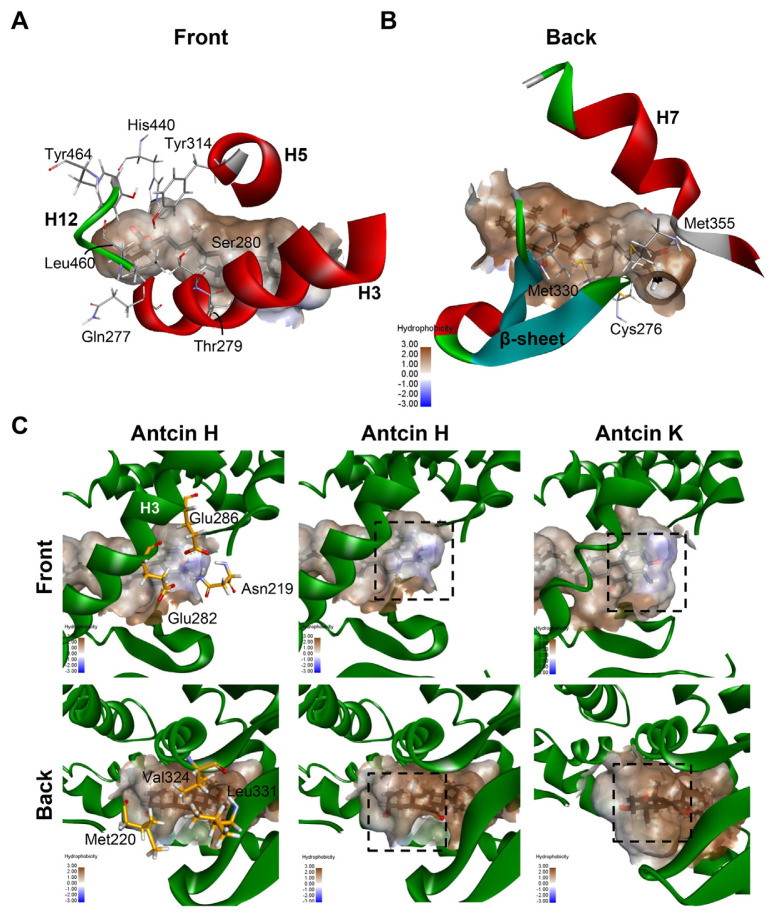
Fig. 6.
Maps of the hydrophobicity at the binding site of PPARα docked with the antcin H and K. (A, B) Ten residues shared by antcins and Wy-14,643 are showed as grey sticks. The bound ligands are shown as black sticks. H3, H5 and H7 of PPARα are shown in ribbon representation. (C) Maps of the hydrophobicity at the binding site of PPARα docked with the antcin H and K. Ligands and receptor residues surrounding ring A of antcins are shown as black and yellow sticks, respectively. Regions surrounding ring A are highlighted in dotted rectangles.

