



Abstract
The canine corpus luteum (CL) is the main source of reproductive steroids during dioestrus in the dog and remains active even in the absence of pregnancy (non‐pregnant dioestrus, physiological pseudopregnancy). Whereas the biological effects of 17β‐oestradiol (E2) in the canine CL remain unclear, the transcriptional availability of oestrogen receptors, ESR1 and ESR2, as well as other modulators of local availability of E2, for example, HSD17B7 (converts oestrone into oestradiol), SULT1E1 (inactivates E2 binding capacity to its own receptors through sulphonation) and STS (reverts E2 sulphonation), were previously detected in the CL of non‐pregnant bitches. The aim of the present work was to evaluate the mRNA amounts of these factors involved in luteal sensitivity and metabolism of E2 in the canine CL during the course of non‐pregnant dioestrus (days 10, 20, 30, 40, 50 and 60 post‐ovulation, n = 5/group) and at different stages of pregnancy (n = 4‐6/group): pre‐implantation (days 8–12), post‐implantation (days 18–25), mid‐gestation (days 35–40) and prepartum luteolysis. During pregnancy, the availability of ESR1, HSD17B7, SULT1E1 and STS decreased from mid‐pregnancy to prepartum luteolysis. The main findings during non‐pregnant dioestrus were as follows: increased ESR2:ESR1 ratio on days 40 and 50 after ovulation, decreasing during luteal regression (day 60); increased STS at day 30 when SULT1E1 levels decreased; increased availability of SULT1E1 transcripts during luteal regression; and decreased amounts of HSD17B7 mRNA in early dioestrus, increasing towards later stages. These results suggest that E2 signalling and biologically active local concentrations could diverge in response to time and pregnancy status of the bitch.
Keywords: 17β‐oestradiol, Corpus luteum, dog (Canis lupus familiaris), non‐pregnant dioestrus, pregnancy
1. INTRODUCTION
In the domestic dog, the reproductive cycle differs from that of other domestic mammals. Among several peculiarities is the lack of a luteolytic signal in the absence of pregnancy, contrasting with the placental PGF2α release prior to parturition (Gram et al., 2013; Hoffmann et al., 1992; Kowalewski, 2014; Kowalewski et al., 2010; Luz et al., 2006). Instead, luteal activity in non‐pregnant bitches is long, frequently surpassing pregnancy itself, and is terminated with a slow luteal regression (reviewed in Kowalewski, 2014). This long life span of the corpus luteum (CL) in the absence of pregnancy results in a physiological pseudopregnancy (non‐pregnant dioestrus). The functional importance of the CL is further highlighted by the absence of steroidogenic activity in the canine placenta (Hoffmann et al., 1994; Nishiyama et al., 1999) rendering the CL the only major source of circulating reproductive steroids during pregnancy. Accordingly, the expression of the respective steroidogenic machinery was confirmed in the canine CL, including the steroidogenic acute regulatory (STAR) protein, 3β‐hydroxysteroid dehydrogenase (HSD3B1) and P450 aromatase (CYP19A1) (Kowalewski et al., 2009; Kowalewski & Hoffmann, 2008; Nishiyama et al., 1999; Papa & Hoffmann, 2011).
E2 is predominantly secreted by granulosa and luteal cells and is involved in a wide range of physiological and pathological mechanisms, with special importance in the reproductive system (reviewed in Fuentes & Silveyra, 2019; Jia et al., 2015). For its production, cholesterol, the substrate for steroid synthesis, needs to be converted into progestogens and androstanes, the latter undergoing aromatization into oestrogens in enzymatic steps mediated by CYP19A1 (Miller, 2017). To obtain the predominant, biologically active E2, the 17β‐hydroxyl group of oestrone (E1), or of the non‐aromatized androstenedione, needs to be oxidized by hydroxysteroid 17β dehydrogenases (HSD17Bs), such as HSD17B1 and HSD17B7 (reviewed in Luu‐The, 2001; Miller, 2017; Stocco et al., 2007).
The effects of E2 are predominantly mediated by its two nuclear receptors, oestrogen receptor α (ERα/NR3A1, encoded by ESR1) and ‐β (ERβ/NR3A2, encoded by ESR2) (Fuentes & Silveyra, 2019; Jia et al., 2015). The availability of oestrogens in target cells can also be modulated through the sulphatase pathway. This pathway involves sulphotransferases, such as the sulphotransferase family 1E member 1 (SULT1E1), that converts active oestrogens into biologically inactive oestrogen sulphates, and the steroid sulphatase (STS) that reverts this mechanism, that is, activates E2 (Purohit et al., 1998; Rizner, 2016; Secky et al., 2013; Song, 2001). In this way, E2 signalling in target tissues can be regulated at different levels, that is, through the modulation of its synthesis or through its sulphonation, thereby affecting E2 availability for specific receptors or, finally, by regulating the local availability of the respective receptors.
Local regulation, that is, autocrine and paracrine, plays an extensive role in maintaining luteal function and involves several factors, including PGE2 and P4. This also applies to the dog (Kowalewski et al., 2013, 2015; Zatta et al., 2017). Regarding E2, it was involved in the release of pituitary prolactin (PRL) in several species (Reymond & Lemarchand‐Béraud, 1976; Stone et al., 1977; Vician et al., 1979). This includes the dog (Jones & Boyns, 1976), where circulating amounts of PRL increase with the progression of the luteal phase, becoming the predominant luteotropic factor in the second half of the canine dioestrus (Okkens et al., 1986, 1990; Onclin & Verstegen, 1997; Onclin et al., 2000). Interestingly, effects of oestrogens in the CL appear to vary among different species, depending on their concentration and local availability. Thus, for example, in the rat and rabbit, oestrogens have luteotropic potential, whereas exogenous administration of oestrogens in rhesus monkeys shortens luteal life span (Hassani et al., 1978; Karsc & Sutton, 1976; Miller & Keyes, 1978; Townson et al., 1996; Tripathy et al., 2016). In the guinea pig, E2 effects appear to be time‐dependent, as administration of oestrogens in early dioestrus induces uterus‐mediated luteolysis, while application after day 9 of dioestrus prolongs luteal function (Illingworth & Perry, 1973). Interestingly, none of this is known for the dog, a species fully dependent on luteal steroids for the maintenance of pregnancy.
During the canine reproductive cycle, the amount of circulating E2 peaks prior to ovulation (1–2 days before the LH surge), decreasing to basal concentrations in early dioestrus (Concannon, 2009; Feldman & Nelson, 2004; Kowalewski, 2018; Onclin et al., 2002; Papa & Hoffmann, 2011). After the time of luteal formation, that is, around day 10 post‐ovulation (p.o.), plasma concentrations of E2 increase again following increased expression of luteal CYP19A1, but never reach values comparable with the preovulatory peak, and follow roughly the secretion patterns observed for P4 (Kowalewski, 2018; Onclin et al., 2002; Papa & Hoffmann, 2011). During luteal regression, the availability of luteal steroids, both P4 and E2, in peripheral plasma, starts to slowly decrease with the passive regression of luteal activity towards basal concentrations; this slow decrease is only interrupted in pregnant animals where, in response to prepartum luteolysis, P4 and E2 return abruptly to basal concentrations (Concannon et al., 1989; Feldman & Nelson, 2004; Hoffmann et al., 1992; Kowalewski, 2014; Onclin et al., 2002). In contrast with what is observed in other species, no pregnancy and/or parturition‐related increase is observed in circulating plasma E2 concentrations (Concannon, 2011; Hoffmann et al., 1994; Holst et al., 2015; Kowalewski, 2018). However, the high variation in circulating E2 observed within and between different reports appears to be related not only to technical limitations (due to the low amounts of circulating E2), but is also associated with individual and/or possible breed divergences (Hoffmann et al., 1992, 1994; Holst et al., 2015; Onclin et al., 2002; Papa & Hoffmann, 2011). At the cellular level, the CL of non‐pregnant bitches is sensitive to E2, as luteal expression of both ERα and ‐β was previously demonstrated (Hoffmann et al., 2004; Papa & Hoffmann, 2011; Tavares Pereira, Gram, et al., 2019). Moreover, in a recent transcriptomic analysis by our group, the expression of HSD17B7 increased after the maturation of the CL in non‐pregnant bitches, and luteal withdrawal of prostaglandins presented stage‐dependent effects in the expression of SULT1E1, suggesting its involvement in local compensatory mechanisms (Tavares Pereira, Graubner, et al., 2019). Such observations suggest the presence of mechanisms in the canine CL that actively regulate its own sensitivity to oestrogens during pseudopregnancy, but nothing is known regarding pregnancy.
Therefore, the aim of the present work was to provide the first insight into the presence of factors involved in E2 signalling and metabolism in the canine CL. For that, we evaluated the availability of transcripts encoding for both nuclear oestrogen receptors (ESR1 and ESR2), HSD17B7 and enzymes involved in the sulphatase pathway of oestrogens (SULT1E1 and STS) in the CL of pregnant and non‐pregnant bitches (i.e. during physiological pseudopregnancy).
2. MATERIALS AND METHODS
2.1. Tissue samples
Luteal tissue was obtained from 50 crossbred and clinically healthy bitches aged 2–8 years by routine ovariohysterectomy at different stages of pregnancy or non‐pregnant dioestrus. All samples used in the present work were derived from previous studies (Kowalewski et al., 2009, 2010; Miskulin Cardoso et al., 2021). Animal experiments were carried out in accordance with animal welfare ethical principles and legislation, and approved by the responsible ethics committees of the Justus‐Liebig University Giessen, Germany (permits no. II 25.3‐19c20‐15c GI 18/14 and VIG3‐19c‐20/15 GI 18,14), of the University of Ankara, Turkey (permits no. Ankara 2006/06 and 2008–25–124) and of the University of São Paulo, Brazil (permit no. 2718/2012). Furthermore, samples from animals that underwent routine ovariohysterectomy at the Section of Small Animal Reproduction, Vetsuisse Faculty, Zurich, Switzerland, were collected after informed consent of the owners was obtained.
Animals were monitored for the onset of spontaneous oestrus by vaginal cytology and/or observation of pro‐oestrus bleeding. Blood samples were then collected every 2 days and the day of ovulation was considered to be when peripheral concentrations of P4 exceeded 5 ng/ml. All samples were collected by routine ovariohysterectomy. Those tissues from non‐pregnant animals (n = 5 animals/group) were sampled on days 10, 20, 30, 40, 50 and 60 post‐ovulation (p.o.). Animals from pregnancy groups were mated 2 days after ovulation (day 0 of pregnancy) and samples were collected at pre‐implantation (days 8–12 of pregnancy, n = 6), post‐implantation (days 18–25 of pregnancy, n = 5), mid‐gestation (days 35–40 of pregnancy, n = 5) stages and at the time of prepartum luteolysis (n = 4). Presence of pregnancy during the pre‐implantation stage was confirmed by embryo flushing. The onset of the parturition cascade in dogs is associated with trophoblast‐derived PGF2α, which induces the cessation of luteal activity (prepartum luteolysis), reflected in a steep decline of circulating P4 levels (reviewed in Kowalewski et al., 2020). Thus, to determine prepartum luteolysis, circulating P4 was measured every 6h from day 58 of pregnancy until the detected levels were below 3ng/ml in three consecutive measurements. Immediately after collection, CLs were dissected from surrounding tissues, immersed in RNALater (Ambion Biotechnology GmbH) for 24h at 4ºC and then stored at −80ºC until extraction of total RNA. For histological analysis, luteal tissue was fixed in 10% neutral phosphate‐buffered formalin for 24 hr, before being washed with PBS, dehydrated and paraffin‐embedded.
2.2. Total RNA isolation, reverse transcription and semi‐quantitative real‐time TaqMan PCR
Total RNA isolation was performed by using the TRIzol reagent (Invitrogen) following the manufacturer's protocol, and RNA concentration and purity was assessed with a NanoDrop 2000 spectrophotometer (Thermo Fisher Scientific AG Reinach). For each sample, 1 μg of total RNA was purified from possible contaminating genomic DNA with the RQ1 RNA‐free DNase kit (Promega) and cDNA was obtained with Superscript III reverse transcriptase (Life Technologies) following the protocol provided by the supplier. cDNA from each sample was pre‐amplified by mixing it with PreAmp Master Mix (Applied Biosystems, Foster City, CA, USA) and pooled TaqMan assays for all target and reference genes, as previously described (Tavares Pereira, Gram, et al., 2019). Table 1 presents the complete list of the self‐designed TaqMan systems and 6‐carboxyfluorescein (6‐FAM) and 6‐carboxytetramethylrhodamine (TAMRA) labelled probes (ordered from Microsynth AG, Balgach, Switzerland), and predesigned commercially available TaqMan assays (Applied Biosystems). The composition of self‐designed TaqMan systems (targeting ESR1, ESR2 and SULT1E1) was previously published and their efficiency was evaluated to ensure each was approximately 100% (Kautz et al., 2014; Tavares Pereira, Graubner, et al., 2019). The availability of mRNA was evaluated by semi‐quantitative real‐time TaqMan PCR using an automated ABI PRISM 7500 Sequence Detection System (Life Technologies) as previously described (Kowalewski et al., 2006, 2011). Briefly, reactions were run with TaqMan Universal Master Mix II (Applied Biosystems) in duplicate in 96‐well optical plates, using autoclaved water or DNAse‐treated RNA (minus‐RT control) instead of cDNA as negative controls. Relative quantification of mRNA expression was performed with the comparative Ct method (ΔΔCt), calibrated to the average expression of all analysed samples and normalized with three reference genes stably expressed in canine reproductive tissue: PTK2, EIF4H and KDM4A (Nowak et al., 2020).
TABLE 1.
List of genes, corresponding TaqMan systems used for semi‐quantitative real‐time PCR and primers used for generation of templates for in situ hybridization (ISH)
| Gene | Name | Accession numbers | Primer and probe sequence for semi‐quantitative real‐time PCR | Product length (bp) | |
|---|---|---|---|---|---|
| ERα/ESR1 | Oestrogen receptor alpha | NM_001286958.1 | Forward | 5′‐CCC ATG GAG GAG ACA AAC CA−3′ | 93 |
| Reverse | 5′‐CCC TGC CTC GGT GAT ATA−3′ | ||||
| TaqMan probe | 5′‐CAC GGG CCC AAC TTC ATC ACA TTC C−3′ | ||||
| ERβ/ESR2 | Oestrogen receptor beta | XM861041 | Forward | 5′‐CCC AGC CCC TTC A−3′ | 78 |
| Reverse | 5′‐AAT CAT ATG CAC GAG TTC CTT GTC−3′ | ||||
| TaqMan probe | 5′‐CCT CCA TGA TGA TGT CCC TGA CC−3′ | ||||
| SULT1E1 | Sulphotransferase family 1E member 1 | MK728829 | Forward | 5′‐AAC AGA TGG CAT CTC CTA GAG TAG TG−3′ | 100 |
| Reverse | 5′‐CGG CAA AGA TAG ATC ACC TTA CAG T−3′ | ||||
| TaqMan probe | 5′‐CCA TCT GCC AGT TGA ACT TCT TCC AGC C−3′ | ||||
| STS | Steroid sulphatase | Applied Biosystems, prod. no. Cf03986178_m1 | 64 | ||
| HSD17B7 | Hydroxysteroid 17β dehydrogenase 7 | Applied Biosystems, prod. no. Cf02657821_m1 | 82 | ||
| PTK2 | Protein tyrosine kinase 2 | Applied Biosystems, prod. no. Cf02684608_m1 | 104 | ||
| EIF4H | Eukaryotic translation initiation factor 4H | Applied Biosystems, prod. no. Cf02713640_m1 | 136 | ||
| KDM4A | Lysine (K)‐specific demethylase 4A | Applied Biosystems, prod. no. Cf02708629_m1 | 96 | ||
| Gene | Accession numbers | Primer sequence for in situ hybridization | Product length (bp) | |
|---|---|---|---|---|
| SULT1E1 | MK728829 | Forward | 5′‐ TGC AAA GAG GGT GAT GTG GAA−3′ | 272 |
| Reverse | 5′‐ GGG TCT GGA TGA GCC GTT AT−3′ | |||
| STS | XM_038449341 | Forward | 5′‐ TGC CCG AGG ACA GAA TCA TC−3′ | 272 |
| Reverse | 5′‐ AAC ATC GAA GAG CAG TGG CG−3′ | |||
| HSD17B7 | XM_014111264.3 | Forward | 5′‐ CGT CTC GCA ATG CAA GGA AA−3′ | 256 |
| Reverse | 5′‐ TAA AGA GGG CGT GCA GAT GA−3′ | |||
2.3. In situ hybridization (ISH)
Localization of the three enzymes involved in E2 metabolism, that is, HSD17B7, SULT1E1 and STS, was performed at the mRNA level with non‐radioactive in situ hybridization (ISH), following previously published protocols (Kowalewski et al., 2010; Kowalewski, Mason, et al., 2006). For the synthesis of cRNA probes, cDNA produced from luteal tissue was amplified with PCR using the canine specific primers (ordered from Microsynth), listed in Table 1. PCR products were separated by electrophoresis in a 2% agarose gel stained with ethidium bromide, purified with the QIAquick Gel Extraction Kit (Qiagen GmBH, Hilden, Germany), ligated into pGEM‐T plasmids (Promega) and cloned in XL1‐Blue Competent Cells (Agilent, Waldbronn, Germany). After monoclonal selection and enrichment, plasmids were isolated with the PureYield Plasmid Miniprep System (Promega), submitted to control digestion with NotI and NcoI restriction enzymes (New England Biolabs, Frankfurt, Germany) and submitted for Sanger sequencing (Microsynth) to ensure the specificity and identify the sense and anti‐sense direction of the products in the plasmids. Plasmids where then linearized either with NotI or NcoI restriction enzyme and digoxigenin (DIG)‐labelled cRNA probes were synthesized with the DIG‐RNA labelling kit (Roche Diagnostics AG, Basel, Switzerland), following the provided protocol. Efficiency of the synthesis of riboprobes was checked by semi‐quantification applying dot‐blot analysis of serial dilutions on positively charged nylon membranes (Roche).
Non‐radioactive ISH was performed on 2μm‐thick sections of formalin‐fixed and paraffin embedded luteal tissue collected from three animals on days 30–35 of pregnancy. After dewaxing with xylol and rehydration in a graded ethanol series, tissue was permeabilized by digestion with 70 µg/ml of proteinase K (Sigma‐Aldrich Chemie GmbH) at 37ºC for 20 min, and post‐fixed with 4% paraformaldehyde. Hybridization was performed overnight at 37ºC in the presence of formamide. The sense probe was used as negative control. For the detection of DIG‐labelled cRNA, tissue was incubated overnight at 4ºC with alkaline phosphatase (AP)‐conjugated sheep anti‐DIG Fab fragments antibody (1:5000, Roche Diagnostics), while endogenous alkaline phosphatases were inhibited with levamisole. Visualization of signals (i.e. of the AP conjugated with anti‐DIG antibody) was performed using 5‐bromo‐4‐chloro‐3‐indolyl phosphate / nitroblue tetrazolium colour development substrate (BCIP/NBT, Roche Diagnostics), yielding blue/purple precipitates at the site of the reaction. After stopping the reaction, slides where mounted with Mowiol 4–88 (Calbiochem, EMD Millipore Inc) and pictures were acquired using a Leica DMRXE light microscope equipped with a Leica Flexacam C1 camera (Leica Microsystems).
2.4. Statistical analysis
The relative amounts of mRNA of most factors did not follow a normal distribution. Thus, data from all target genes were normalized with a logarithmic transformation before statistical analysis. Numerical results are presented as geometric mean ± geometric standard deviation (SD). Time‐related changes in the mRNA levels of investigated target genes, as well as in the ratio ESR2:ESR1, were evaluated with the Kruskal–Wallis’ non‐parametric ANOVA followed, if it reported p < .05, by the Tukey–Kramer multiple comparisons test, using the software GraphPad3 (GraphPad Software Inc.). p < .05 was considered as significant.
3. RESULTS
The availability of all factors investigated was detectable in all analysed luteal samples from pregnant and non‐pregnant animals. Levels of ESR1 changed in a time‐dependent manner in the CL of pregnant animals (p < .0001, Figure 1a). Luteal availability of ESR1 transcripts increased gradually from pre‐implantation (Pre‐Imp) towards mid‐gestation (p < .05), when the highest mRNA amounts were observed, and then decreased significantly during prepartum luteolysis (p < .001). However, during non‐pregnant dioestrus, luteal expression of ESR1 displayed high individual variability, and no significant changes were observed (p > .05, Figure 1b). An inverse situation was observed for ESR2, which presented no stage‐dependent changes in its mRNA abundance during pregnancy (p > .05, Figure 1c), but did in the absence of pregnancy (p < .05, Figure 1d). In fact, non‐pregnant dioestrus was marked by a significantly higher availability of ESR2 at day 40 post‐ovulation (p.o.) than on days 10 or 20 (p < .05). Following this, the ratio between the transcriptional availability of both oestrogen receptors was assessed and was found to be affected by the progression of dioestrus both during pregnancy (p < .01, Figure 1e) and non‐pregnant luteal stage (p < .001, Figure 1f). In samples from pregnant animals, the ESR2:ESR1 ratio significantly decreased towards mid‐gestation (p < .05), changing later and increasing during prepartum luteolysis (p < .001). In the absence of pregnancy, the ratio between ESR2 and ESR1 transcript levels was lower at day 10 p.o. than on days 30, 40 and 50 (p < .05). Furthermore, this ratio was also significantly higher on day 40 than days 20 and 60 p.o. (p < .05).
FIGURE 1.
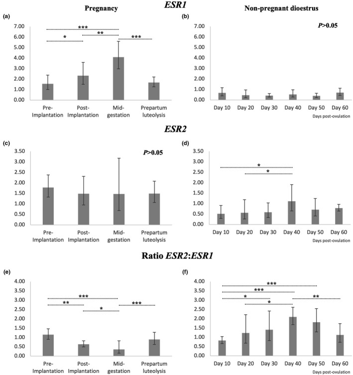
Relative mRNA abundance of ESR1, ESR2 and the ESR2/ESR1 mRNA ratio determined by semi‐quantitative real‐time (TaqMan) PCR in the canine CL at selected stages of pregnancy and pseudopregnancy. Samples from pregnant animals were collected pre‐implantation (between days 8–12), post‐implantation (days 18–25) and at mid‐gestation (days 35–40) of pregnancy, or at the time of prepartum luteolysis. Time‐points from non‐pregnant dioestrus refer to days post‐ovulation. Data are presented as geometric mean ± geometrical standard deviation. In the case of the one‐way ANOVA reporting p < .05, analysis was followed by a Tukey–Kramer multiple comparisons post‐test. Bars with asterisks differ at: * = p < .05, ** = p < .01, *** = p < .001
The mRNA levels encoding for the three enzymes involved in the regulation of E2 availability changed significantly in the CL during pregnancy (p < .0001 for all factors) and non‐pregnant dioestrus (p < .0001 for SULT1E1 and p < .001 for HSD17B7 and STS, Figure 2). Neither HSD17B7 nor STS varied significantly during pre‐implantation and post‐implantation, but the mRNA amounts of both decreased in later stages of pregnancy, being the lowest during prepartum luteolysis (p < .05, Figure 2a and e respectively). In non‐pregnant animals, luteal abundance of HSD17B7 was the lowest on day 10 p.o., and was also decreased on day 20 when compared with days 40 and 60 (p < .05, Figure 2b), apparently showing a different dioestrus‐related expression pattern than during pregnancy. The absence of pregnancy was also associated with a higher STS mRNA levels at day 30 than on days 10, 20 and 50 p.o. (p < .05, Figure 2f). The luteal availability of SULT1E1 during pregnancy increased from pre‐implantation to post‐implantation and mid‐gestation (p < .01, Figure 2c). However, similarly to HSD17B7 and STS, it was decreased at the time of luteolysis (p < .01, Figure 2c). Conversely, in the absence of pregnancy, the levels of transcripts encoding for SULT1E1 decreased from day 10 p.o. to days 20 and 30 (p < .01, Figure 2d), but were significantly increased during late dioestrus, that is, days 50 and 60 p.o., when compared with all previously analysed time‐points (p < .001, Figure 2d). The localization of HSD17B7, SULT1E1 and STS was performed at the mRNA level by ISH. Positive signals for all enzymes were mainly detected in luteal cells (Figure 2g–i). Furthermore, positive staining against HSD17B7 could also be observed in endothelial cells (Figure 2g). There was no staining observed in negative controls (sense riboprobes).
FIGURE 2.
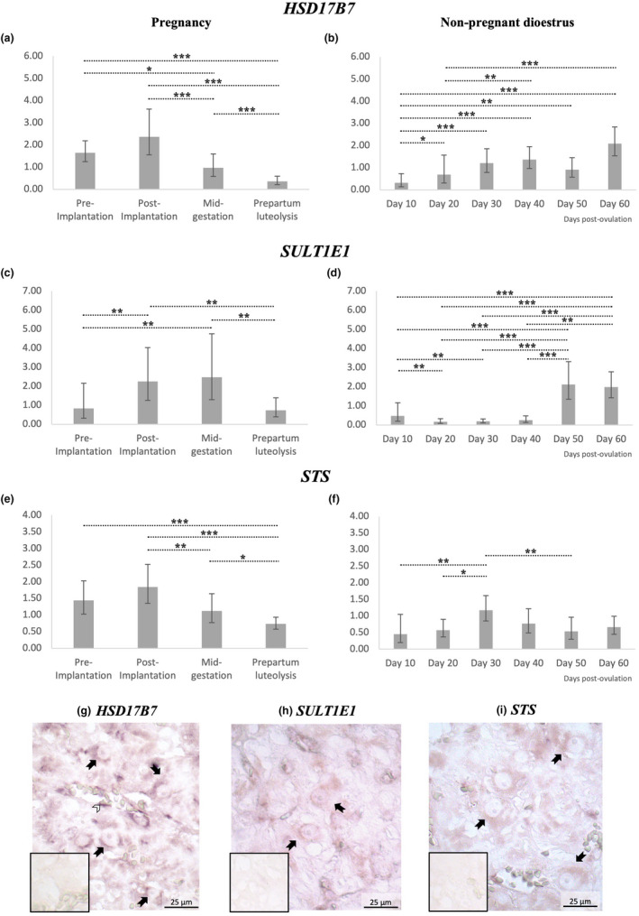
Relative abundance and localization of HSD17B7, SULT1E1 and STS mRNA in the canine CL. (a‐f) Relative mRNA abundance was determined by semi‐quantitative real‐time (TaqMan) PCR. Samples from pregnant animals were collected pre‐implantation (between days 8–12), post‐implantation (days 18–25) and at mid‐gestation (days 35–40) of pregnancy, or at the time of prepartum luteolysis. Time‐points from non‐pregnant dioestrus refer to days post‐ovulation. Data are presented as geometric mean ± geometrical standard deviation. In the case of the one‐way ANOVA reporting p < .05, analysis was followed by a Tukey–Kramer multiple comparisons post‐test. Bars with asterisks differ at: * = p < .05, ** = p < .01, *** = p < .001. (g–i) The luteal localization of transcripts encoding for HSD17B7, SULT1E1 and STS was performed in mid‐pregnant dogs by in situ hybridization (ISH). Positive signals for all factors were mainly observed in luteal cells (closed arrows). mRNA encoding for HSD17B7 was further detected in endothelial cells (open arrowheads). No staining was observed in the negative controls (sense probe; insets in figures, at the same magnification)
4. DISCUSSION
Based on the luteal presence of oestrogen receptors described previously (Hoffmann, Büsges, Engel, et al., 2004; Papa & Hoffmann, 2011) and further elaborated in the present study, it appears that E2 conveys autocrine/paracrine effects in the canine CL. Considering the similarity between circulating profiles of P4 and E2 (Hoffmann et al., 1992; Onclin et al., 2002), possible luteotropic effects were proposed for E2 within the canine CL (Papa & Kowalewski, 2020). With all of this in mind, we felt prompted to investigate the expression of factors involved in regulating metabolism and possible signalling pathways of E2 in the canine CL. Accordingly, here, we report for the first time the luteal levels of transcripts encoding for ESR1, ESR2, HSD17B7, SULT1E1 and STS in pregnant bitches. We also present the mRNA expression profiles of these factors in the CL throughout non‐pregnant dioestrus (physiological pseudopregnancy). The samples included the following developmental stages of the canine CL: developing CL (day 10 of non‐pregnant dioestrus and pre‐implantation stage, that comprises days 8–12 of pregnancy), mature CL (days 20 and 30 of non‐pregnant dioestrus and post‐implantation stage, that comprises days 18–25 of pregnancy), early regressing CL (day 40 of non‐pregnant dioestrus and samples representing mid‐gestation, that comprises days 35–40 of pregnancy), late luteal regression (day 60 of pseudopregnancy) and prepartum luteolysis (Hoffmann et al., 2004; Kowalewski, 2014). There were no canine‐specific and/or cross‐reacting antibodies suitable for the study that would allow investigations at the protein level. Thus, clearly, before any final functional conclusions can be drawn, the respective information needs to be provided.
Nevertheless, time‐dependent changes in the availability of ESR1 transcripts were observed during pregnancy, increasing towards mid‐term, whereas ESR2 amounts remained unaffected at selected time points during pregnancy. In non‐pregnant animals, no significant changes of the transcript levels of ESR1 were observed in response to the passage of time. On the other hand, there were higher amounts of ESR2 transcripts on day 40 p.o., when compared with early luteal stages (days 10 and 20), resulting in a higher ESR2/ESR1 ratio. Apparently, the results we obtained for CL of non‐pregnant dogs are in contrast with previous observations showing time‐dependent effects, for example, in the increased mRNA availability of ESR1 in mid‐dioestrus (Papa & Hoffmann, 2011; Tavares Pereira, Gram, et al., 2019). A possible explanation could be in the strongly varying circulating E2 concentrations regulating the local availability of its own receptors in a self‐regulatory feedback loop, possibly similar to what is observed, for example, with P4, which appears to regulate the expression of its own nuclear receptor (Papa & Hoffmann, 2011). Moreover, the different time points of tissue collection evaluated in the present and previous works (Papa & Hoffmann, 2011; Tavares Pereira, Gram, et al., 2019) could further explain some discrepancies in the pattern of the herein and previously observed intraluteal ESR1 mRNA availability.
An interesting observation from the present study arises from the changes in the ESR2:ESR1 ratio. The role of oestrogen receptors in the CL is still poorly understood, although some insights into possible functions are provided from other biological systems. Thus, for example, in the murine uterus, ERα appeared to be related to increased proliferative activity, whereas ERβ showed antiproliferative effects (Weihua et al., 2000). These opposing effects were also observed in other healthy and neoplastic tissues, with ERα being more frequently associated with the promotion of cell growth and proliferation and ERβ blocking such mechanisms, probably associated with a differential regulation of factors involved in cell cycle modulation like, for example, cyclin D1 (Fuentes & Silveyra, 2019; Liu et al., 2002; Lucas et al., 2014). Additionally, activated ERα and ERβ may form heterodimers that evoke ERα‐related effects in the transcription of target genes (Fuentes & Silveyra, 2019; Li et al., 2004). The importance of the ESR2:ESR1 ratio for the functionality of the canine CL has been indicated recently, with its variation possibly being implicated in the regulation of proliferative and/or metabolic mechanisms (Papa & Kowalewski, 2020). The decreased ESR2:ESR1 ratio observed here during mid‐pregnancy suggested a dominance of ERα signalling over ERβ during the maintenance of pregnancy in the dog that, apparently, is later reverted at the time of luteolysis. This pattern highly contrasted with non‐pregnant dioestrus, where the abundance of ESR2 transcripts was higher than ESR1 transcripts in mid‐dioestrus, but not in the earlier stages or during late luteal regression. While protein availability and functional studies are still required for a full understanding of E2 signalling in the canine CL, the present results suggest the occurrence of changes in the luteal sensitivity to oestrogens induced by pregnancy in the dog, by affecting not only the individual availability of each oestrogen receptor, but mainly the relative proportions of ESR1 and ESR2.
The local availability of E2 is, in addition to the presence of oestrogen receptors, essential for the signalling of this steroid hormone. With this in mind, we evaluated the abundance of transcripts encoding for enzymes involved in the production of E2 (HSD17B7) or modulating the capacity of E2 to bind to its own receptors (SULT1E1 and STS). The lowest availability of HSD17B7 during pseudopregnancy occurred early, on days 10 and 20 p.o., in agreement with our previous observations (Tavares Pereira, Graubner, et al., 2019). HSD17B7 is the sole HSD17B enzyme in the corpus luteum of the rat, and its luteal expression has also been described in species such as human and ruminants (Nokelainen et al., 1998; Parmer et al., 1992). It can convert oestrone into oestradiol, but not androstenedione into testosterone (Luu‐The, 2001; Marijanovic et al., 2003; Nokelainen et al., 1998; Peltoketo et al., 1999; Törn et al., 2003). Thus, the possible increased availability of HSD17B7 after the early luteal stage, associated with the clearly abundant presence of CYP19A1 observed in mid‐dioestrus (Papa & Hoffmann, 2011), implies the contribution of HSD17B7 in the production of E2 during luteal maturation. Furthermore, the abundance of the inactivator of E2 binding‐capacity to its receptors, SULT1E1, appears to be increased during late dioestrus, at days 50 and 60, implying its involvement in the functional withdrawal of E2 in regressing CL. This possible endocrine mechanism was also implied from previous transcriptomic studies, in which the increased presence of SULT1E1 was reported during luteal regression (Zatta et al., 2017). This mechanism appears to be coordinated with the expression of STS, showing opposite mRNA availability patterns, with the highest levels on day 30 p.o., when the presence of SULT1E1 was decreased, and lowered towards day 50 p.o.. The overall gestational mRNA expression patterns of HSD17B7, SULT1E1 and STS seemed to differ from those observed at non‐pregnant dioestrus. While presenting higher availability in earlier pregnancy stages, the amount of mRNA of all three factors was strongly decreased during the cessation of luteal function, that is, at prepartum luteolysis. Taking into consideration that the natural or antigestagen‐mediated withdrawal of P4 leads to active termination of luteal function, associated with strong apoptotic activity (Galac et al., 2000; Kowalewski, 2014; Zatta et al., 2017), the termination of steroidogenic activity in the CL during prepartum luteolysis is not surprising. This situation contrasts with the passive luteal regression devoid of apoptotic activity observed in non‐pregnant dogs (Hoffmann, Büsges, Engel, et al., 2004; Sonnack, 2009). Indeed, the ongoing luteal degeneration, and shortage in the provision of steroidogenic substrate, were indicated as the rate‐limiting steps and the main cause of the functional withdrawal of luteal function in pseudopregnant dogs (Hoffmann, Büsges, Engel, et al., 2004; Kowalewski & Hoffmann, 2008). In this context, the decreased expression of HSD3B1 was considered secondary (Kowalewski & Hoffmann, 2008). Thus, the physiological differences in mechanisms involved in the cessation of luteal function between pregnant and non‐pregnant dogs need to be taken into consideration when interpreting the data presented here. Accordingly, otherwise, luteal development and maintenance in both pregnant and non‐pregnant dogs were associated with elevated levels of HSD17B7 and STS, involved in regulating the local availability of E2. Last but not least, when the local, that is, intraluteal, activity of oestrogens is considered, the possible role of E2 in regulating the expression of the PRL receptor (PRLR) should be taken into account. Such a mechanism has been shown in other species and organs, for example, in the brain of rats or human pituitary adenoma (DeVito et al., 1992; Shamgochian et al., 1995; Xiao et al., 2020).
Finally, the expression of HSD17B7, SULT1E1 and STS was localized in the canine CL by ISH. Samples from animals between days 30 and 35 of gestation (mid‐gestation) were selected, because relatively high transcriptional availability of all factors was observed during that time of gestation, ensuring required detection limits that are frequently limiting for the application of ISH. Moreover, this time period relates to a fully developed but not yet regressing CL. Complementing previous descriptions of the localization of ERs in lutein and non‐lutein cells (Hoffmann, Büsges, Engel, et al., 2004; Papa & Hoffmann, 2011), HSD17B7 expression was observed in both lutein and endothelial cells. The latter observation, that is, the presence of HSD17B7 in endothelial cells of the CL, seems to be an interesting finding. The role of HSD17B7 in angiogenesis was previously suggested from observation in mice, where specific HSD17B7 knockout resulted in embryonic death, associated with decreased vascularization of yolk sac and altered differentiation of cardiac tissue (Jokela et al., 2010). Clearly, however, the functional role of HSD17B7 in the endothelium still needs further clarification. SULT1E1 and STS signals were observed in lutein cells, similar to other steroidogenic factors, that is, STAR, HSD3B1 and P450 aromatase (Kowalewski & Hoffmann, 2008; Kowalewski, Mason, et al., 2006; Papa & Hoffmann, 2011). Considering this localization, it appears that, in addition to be the main source of reproductive steroids during dioestrus, lutein cells, and to some extend also luteal endothelial cells, might be further involved in the activation/inactivation of E2 produced by the CL. This implies not only auto‐ and paracrine relevance, but possibly also a systemic significance.
5. CONCLUSION
The signalling and, therefore, possible biological effects of E2 may depend on the reproductive status of a bitch and the functional status of the CL (late luteal regression versus. prepartum luteolysis). ERβ‐mediated signalling (which is possibly anti‐proliferative) appears to be more prevalent during mid‐dioestrus. At that time STS is also increased, and an active withdrawal of the local signalling of E2 in the canine CL seems to be suggested by the increased availability of transcripts encoding for SULT1E1 during luteal regression. In contrast, pregnancy appears to be associated with increased ERα signalling during luteal maintenance with active luteolysis changing the ratio toward the dominance of ERβ. Yet, the exact role of E2 in the canine CL needs further clarification, involving post‐transcriptional regulatory mechanisms and functional studies. That said, the most important findings from the present study suggest an active metabolism of E2 in the canine CL, possibly involving auto‐ and paracrine mechanisms. Finally, clearly, any conclusions regarding the physiological function of E2 in the canine CL should not be drawn based on its strongly varying circulating amounts.
CONFLICT OF INTEREST
The authors declare that they have no conflicts of interest.
AUTHOR CONTRIBUTIONS
MTP developed the concept of the present study, was involved in experimental design, generating, analysing and interpreting data and drafting the manuscript. PP, IMR and SA were involved in the collection of tissue material, knowledge transfer, critical discussion of data and revision of the manuscript. MPK was involved in developing and supervising the project and interpretation of data, drafting and revision of the manuscript. All authors read and approved the final manuscript.
ACKNOWLEDGEMENTS
The authors are thankful to Dr. Sharon Mortimer for the careful editing of the manuscript. The technical expertise and contributions of Ricardo Fernandez Rubia and Kirstin Skaar are greatly appreciated. Part of the laboratory work was performed using the logistics at the Center for Clinical Studies (ZKS), Vetsuisse Faculty, University of Zurich. Open access funding provided by Universitat Zurich. WOA Institution: Universitat Zurich Blended DEAL: CSAL.
Tavares Pereira, M. , Papa, P. , Reichler, I. M. , Aslan, S. , & Kowalewski, M. P. (2022). Luteal expression of factors involved in the metabolism and sensitivity to oestrogens in the dog during pregnancy and in non‐pregnant cycle. Reproduction in Domestic Animals, 57, 86–97. 10.1111/rda.14032
Funding information
The present work was supported by the Swiss National Science Foundation (SNSF) research grant number 31003A_182481.
DATA AVAILABILITY
The data that support the findings of this study are available from the corresponding author upon reasonable request.
REFERENCES
- Concannon, P. W. (2009). Endocrinologic control of normal canine ovarian function. Reproduction in Domestic Animals, 44(Suppl 2), 3–15. 10.1111/j.1439-0531.2009.01414.x [DOI] [PubMed] [Google Scholar]
- Concannon, P. W. (2011). Reproductive cycles of the domestic bitch. Animal Reproduction Science, 124(3–4), 200–210. 10.1016/j.anireprosci.2010.08.028 [DOI] [PubMed] [Google Scholar]
- Concannon, P. W. , McCann, J. P. , & Temple, M. (1989). Biology and endocrinology of ovulation, pregnancy and parturition in the dog. Journal of Reproduction and Fertility. Supplement, 39(0449–3087), 3–25. [PubMed] [Google Scholar]
- DeVito, W. J. , Avakian, C. , Stone, S. , & Ace, C. I. (1992). Estradiol increases prolactin synthesis and prolactin messenger ribonucleic acid in selected brain regions in the hypophysectomized female rat. Endocrinology, 131(5), 2154–2160. 10.1210/endo.131.5.1425416 [DOI] [PubMed] [Google Scholar]
- Feldman, E. C. , & Nelson, R. W. (2004). Ovarian cycle and vaginal cytology. In Feldman E. C., & Nelson R. W. (Eds.), Canine and feline endocrinology and reproduction (3rd ed.). Saunders. [Google Scholar]
- Fuentes, N. , & Silveyra, P. (2019). Estrogen receptor signaling mechanisms. Advances in Protein Chemistry and Structural Biology, 116, 135–170. 10.1016/bs.apcsb.2019.01.001 [DOI] [PMC free article] [PubMed] [Google Scholar]
- Galac, S. , Kooistra, H. S. , Butinar, J. , Bevers, M. M. , Dieleman, S. J. , Voorhout, G. , & Okkens, A. C. (2000). Termination of mid‐gestation pregnancy in bitches with aglepristone, a progesterone receptor antagonist. Theriogenology, 53(4), 941–950. 10.1016/S0093-691X(00)00241-7 [DOI] [PubMed] [Google Scholar]
- Gram, A. , Buchler, U. , Boos, A. , Hoffmann, B. , & Kowalewski, M. P. (2013). Biosynthesis and degradation of canine placental prostaglandins: Prepartum changes in expression and function of prostaglandin F2alpha‐synthase (PGFS, AKR1C3) and 15‐hydroxyprostaglandin dehydrogenase (HPGD). Biology of Reproduction, 89(1), 2. 10.1095/biolreprod.113.109918 [DOI] [PubMed] [Google Scholar]
- Hassani, M. , Roos, J. , & Aron, C. (1978). The adrenal cortex and the luteotrophic action of estrogens during the estrous cycle in the rat. Endokrinologie, 72(1), 43–50. [PubMed] [Google Scholar]
- Hoffmann, B. , Büsges, F. , & Baumgärtne, W. (2004). Immunohistochemical detection of CD4‐, CD8‐ and MHCII‐expressing immune cells and endoglin in the canine corpus luteum at different stages of dioestrus. Reproduction in Domestic Animals, 39(6), 391–395. 10.1111/j.1439-0531.2004.00520.x [DOI] [PubMed] [Google Scholar]
- Hoffmann, B. , Büsges, F. , Engel, E. , Kowalewski, M. P. , & Papa, P. C. (2004). Regulation of corpus luteum‐function in the bitch. Reproduction in Domestic Animals, 39(4), 232–240. 10.1111/j.1439-0531.2004.00508.x [DOI] [PubMed] [Google Scholar]
- Hoffmann, B. , Höveler, R. , Hasan, S. H. , & Failing, K. (1992). Ovarian and pituitary function in dogs after hysterectomy. Journal of Reproduction and Fertility, 96(2), 837–845. 10.1530/jrf.0.0960837 [DOI] [PubMed] [Google Scholar]
- Hoffmann, B. , Höveler, R. , Nohr, B. , & Hasan, S. H. (1994). Investigations on hormonal changes around parturition in the dog and the occurrence of pregnancy‐specific non conjugated oestrogens. Experimental and Clinical Endocrinology & Diabetes, 102(3), 185–189. 10.1055/s-0029-1211280 [DOI] [PubMed] [Google Scholar]
- Holst, B. S. , Kushnir, M. M. , & Bergquist, J. (2015). Liquid chromatography‐tandem mass spectrometry (LC‐MS/MS) for analysis of endogenous steroids in the luteal phase and early pregnancy in dogs: A pilot study. Veterinary Clinical Pathology, 44(4), 552–558. 10.1111/vcp.12301 [DOI] [PubMed] [Google Scholar]
- Illingworth, D. V. , & Perry, J. S. (1973). Effects of oestrogen, administered early or late in the oestrous cycle, upon the survival and regression of the corpus luteum of the guinea‐pig. Journal of Reproduction and Fertility, 33(3), 457–467. 10.1530/jrf.0.0330457 [DOI] [PubMed] [Google Scholar]
- Jia, M. , Dahlman‐Wright, K. , & Gustafsson, J. A. (2015). Estrogen receptor alpha and beta in health and disease. Best Practice & Research Clinical Endocrinology & Metabolism, 29(4), 557–568. 10.1016/j.beem.2015.04.008 [DOI] [PubMed] [Google Scholar]
- Jokela, H. , Rantakari, P. , Lamminen, T. , Strauss, L. , Ola, R. , Mutka, A. L. , Gylling, H. , Miettinen, T. , Pakarinen, P. , Sainio, K. , & Poutanen, M. (2010). Hydroxysteroid (17beta) dehydrogenase 7 activity is essential for fetal de novo cholesterol synthesis and for neuroectodermal survival and cardiovascular differentiation in early mouse embryos. Endocrinology, 151(4), 1884–1892. 10.1210/en.2009-0928 [DOI] [PubMed] [Google Scholar]
- Jones, G. E. , & Boyns, A. R. (1976). Oestradiol stimulation of prolactin release from canine pituitary in culture. Acta Endocrinol (Copenh), 82(4), 706–709. 10.1530/acta.0.0820706 [DOI] [PubMed] [Google Scholar]
- Karsc, F. J. , & Sutton, G. P. (1976). An intra‐ovarian site for the luteolytic action of estrogen in the rhesus monkey. Endocrinology, 98(3), 553–561. 10.1210/endo-98-3-553 [DOI] [PubMed] [Google Scholar]
- Kautz, E. , Gram, A. , Aslan, S. , Ay, S. S. , Selcuk, M. , Kanca, H. , Koldas, E. , Akal, E. , Karakas, K. , Findik, M. , Boos, A. , & Kowalewski, M. P. (2014). Expression of genes involved in the embryo‐maternal interaction in the early‐pregnant canine uterus. Reproduction, 147(5), 703–717. 10.1530/REP-13-0648 [DOI] [PubMed] [Google Scholar]
- Kowalewski, M. P. (2014). Luteal regression vs. prepartum luteolysis: Regulatory mechanisms governing canine corpus luteum function. Reproductive Biology, 14(2), 89–102. 10.1016/j.repbio.2013.11.004 [DOI] [PubMed] [Google Scholar]
- Kowalewski, M. P. (2018). Selected comparative aspects of canine female reproductive physiology. In Skinner M. K. (ed.), Encyclopedia of reproduction (2nd ed., pp. 682‐691). Academic Press. [Google Scholar]
- Kowalewski, M. P. , Beceriklisoy, H. B. , Aslan, S. , Agaoglu, A. R. , & Hoffmann, B. (2009). Time related changes in luteal prostaglandin synthesis and steroidogenic capacity during pregnancy, normal and antiprogestin induced luteolysis in the bitch. Animal Reproduction Science, 116(1–2), 129–138. 10.1016/j.anireprosci.2008.12.011 [DOI] [PubMed] [Google Scholar]
- Kowalewski, M. P. , Beceriklisoy, H. B. , Pfarrer, C. , Aslan, S. , Kindahl, H. , Kucukaslan, I. , & Hoffmann, B. (2010). Canine placenta: A source of prepartal prostaglandins during normal and antiprogestin‐induced parturition. Reproduction, 139(3), 655–664. 10.1530/REP-09-0140 [DOI] [PubMed] [Google Scholar]
- Kowalewski, M. P. , Fox, B. , Gram, A. , Boos, A. , & Reichler, I. (2013). Prostaglandin E2 functions as a luteotrophic factor in the dog. Reproduction, 145(3), 213–226. 10.1530/REP-12-0419 [DOI] [PubMed] [Google Scholar]
- Kowalewski, M. P. , & Hoffmann, B. (2008). Molecular cloning and expression of StAR protein in the canine corpus luteum during dioestrus. Experimental and Clinical Endocrinology & Diabetes, 116(3), 158–161. 10.1055/s-2007-992121 [DOI] [PubMed] [Google Scholar]
- Kowalewski, M. P. , Ihle, S. , Siemieniuch, M. J. , Gram, A. , Boos, A. , Zdunczyk, S. , Fingerhut, J. , Hoffmann, B. , Schuler, G. , Jurczak, A. , Domoslawska, A. , & Janowski, T. (2015). Formation of the early canine CL and the role of prostaglandin E2 (PGE2) in regulation of its function: An in vivo approach. Theriogenology, 83(6), 1038–1047. 10.1016/j.theriogenology.2014.12.006 [DOI] [PubMed] [Google Scholar]
- Kowalewski, M. P. , Mason, J. I. , Howie, A. F. , Morley, S. D. , Schuler, G. , & Hoffmann, B. (2006). Characterization of the canine 3beta‐hydroxysteroid dehydrogenase and its expression in the corpus luteum during diestrus. Journal of Steroid Biochemistry and Molecular Biology, 101(4–5), 254–262. 10.1016/j.jsbmb.2006.06.029 [DOI] [PubMed] [Google Scholar]
- Kowalewski, M. P. , Meyer, A. , Hoffmann, B. , Aslan, S. , & Boos, A. (2011). Expression and functional implications of peroxisome proliferator‐activated receptor gamma (PPARgamma) in canine reproductive tissues during normal pregnancy and parturition and at antiprogestin induced abortion. Theriogenology, 75(5), 877–886. 10.1016/j.theriogenology.2010.10.030 [DOI] [PubMed] [Google Scholar]
- Kowalewski, M. P. , Schuler, G. , Taubert, A. , Engel, E. , & Hoffmann, B. (2006). Expression of cyclooxygenase 1 and 2 in the canine corpus luteum during diestrus. Theriogenology, 66(6–7), 1423–1430. 10.1016/j.theriogenology.2006.01.039 [DOI] [PubMed] [Google Scholar]
- Kowalewski, M. P. , Tavares Pereira, M. , & Kazemian, A. (2020). Canine conceptus‐maternal communication during maintenance and termination of pregnancy, including the role of species‐specific decidualization. Theriogenology, 150, 329–338. 10.1016/j.theriogenology.2020.01.082 [DOI] [PubMed] [Google Scholar]
- Li, X. , Huang, J. , Yi, P. , Bambara, R. A. , Hilf, R. , & Muyan, M. (2004). Single‐chain estrogen receptors (ERs) reveal that the ERalpha/beta heterodimer emulates functions of the ERalpha dimer in genomic estrogen signaling pathways. Molecular and Cellular Biology, 24(17), 7681–7694. 10.1128/MCB.24.17.7681-7694.2004 [DOI] [PMC free article] [PubMed] [Google Scholar]
- Liu, M. M. , Albanese, C. , Anderson, C. M. , Hilty, K. , Webb, P. , Uht, R. M. , Price, R. H. Jr , Pestell, R. G. , & Kushner, P. J. (2002). Opposing action of estrogen receptors alpha and beta on cyclin D1 gene expression. Journal of Biological Chemistry, 277(27), 24353–24360. 10.1074/jbc.M201829200 [DOI] [PubMed] [Google Scholar]
- Lucas, T. F. G. , Lazari, M. F. M. , & Porto, C. S. (2014). Differential role of the estrogen receptors ESR1 and ESR2 on the regulation of proteins involved with proliferation and differentiation of Sertoli cells from 15‐day‐old rats. Molecular and Cellular Endocrinology, 382(1), 84–96. 10.1016/j.mce.2013.09.015 [DOI] [PubMed] [Google Scholar]
- Luu‐The, V. (2001). Analysis and characteristics of multiple types of human 17beta‐hydroxysteroid dehydrogenase. Journal of Steroid Biochemistry and Molecular Biology, 76(1–5), 143–151. 10.1016/s0960-0760(00)00155-2 [DOI] [PubMed] [Google Scholar]
- Luz, M. R. , Bertan, C. M. , Binelli, M. , & Lopes, M. D. (2006). Plasma concentrations of 13,14‐dihydro‐15‐keto prostaglandin F2‐alpha (PGFM), progesterone and estradiol in pregnant and nonpregnant diestrus cross‐bred bitches. Theriogenology, 66(6–7), 1436–1441. 10.1016/j.theriogenology.2006.01.036 [DOI] [PubMed] [Google Scholar]
- Marijanovic, Z. , Laubner, D. , Moller, G. , Gege, C. , Husen, B. , Adamski, J. , & Breitling, R. (2003). Closing the gap: Identification of human 3‐ketosteroid reductase, the last unknown enzyme of mammalian cholesterol biosynthesis. Molecular Endocrinology, 17(9), 1715–1725. 10.1210/me.2002-0436 [DOI] [PubMed] [Google Scholar]
- Miller, J. B. , & Keyes, P. L. (1978). Transition of the rabbit corpus luteum to estrogen dependence during early luteal development. Endocrinology, 102(1), 31–38. 10.1210/endo-102-1-31 [DOI] [PubMed] [Google Scholar]
- Miller, W. L. (2017). Steroidogenesis: Unanswered questions. Trends in Endocrinology and Metabolism, 28(11), 771–793. 10.1016/j.tem.2017.09.002 [DOI] [PubMed] [Google Scholar]
- Miskulin Cardoso, A. P. M. , Tavares Pereira, M. , Silva, R. D. , Sousa, L. M. , Giometti, I. C. , Kowalewski, M. P. , & Papa, P. C. (2021). Global transcriptome analysis implicates cholesterol availability in the regulation of canine cyclic luteal function. General and Comparative Endocrinology, 307, 113759. 10.1016/j.ygcen.2021.113759 [DOI] [PubMed] [Google Scholar]
- Nishiyama, T. , Tsumagari, S. , Ito, M. , Kimura, J. , Watanabe, G. , Taya, K. , & Takeishi, M. (1999). Immunohistochemical study of steroidogenic enzymes in the ovary and placenta during pregnancy in the dog. Anatomia, Histologia, Embryologia: Journal of Veterinary Medicine Series C, 28(2), 125–129. 10.1046/j.1439-0264.1999.00170.x [DOI] [PubMed] [Google Scholar]
- Nokelainen, P. , Peltoketo, H. , Vihko, R. , & Vihko, P. (1998). Expression cloning of a novel estrogenic mouse 17 beta‐hydroxysteroid dehydrogenase/17‐ketosteroid reductase (m17HSD7), previously described as a prolactin receptor‐associated protein (PRAP) in rat. Molecular Endocrinology, 12(7), 1048–1059. 10.1210/mend.12.7.0134 [DOI] [PubMed] [Google Scholar]
- Nowak, M. , Aslan, S. , & Kowalewski, M. P. (2020). Determination of novel reference genes for improving gene expression data normalization in selected canine reproductive tissues ‐ A multistudy analysis. BMC Veterinary Research, 16(1), 440. 10.1186/s12917-020-02635-6 [DOI] [PMC free article] [PubMed] [Google Scholar]
- Okkens, A. C. , Bevers, M. M. , Dieleman, S. J. , & Willemse, A. H. (1990). Evidence for prolactin as the main luteotrophic factor in the cyclic dog. The Veterinary Quarterly, 12(4), 193–201. 10.1080/01652176.1990.9694266 [DOI] [PubMed] [Google Scholar]
- Okkens, A. C. , Dieleman, S. J. , Bevers, M. M. , Lubberink, A. A. M. E. , & Willemse, A. H. (1986). Influence of hypophysectomy on the lifespan of the corpus luteum in the cyclic dog. Journal of Reproduction and Fertility, 77(1), 187–192. 10.1530/jrf.0.0770187 [DOI] [PubMed] [Google Scholar]
- Onclin, K. , Murphy, B. , & Verstegen, J. P. (2002). Comparisons of estradiol, LH and FSH patterns in pregnant and nonpregnant beagle bitches. Theriogenology, 57(8), 1957–1972. 10.1016/s0093-691x(02)00644-1 [DOI] [PubMed] [Google Scholar]
- Onclin, K. , & Verstegen, J. P. (1997). In vivo investigation of luteal function in dogs: Effects of cabergoline, a dopamine agonist, and prolactin on progesterone secretion during mid‐pregnancy and ‐diestrus. Domestic Animal Endocrinology, 14(1), 25–38. 10.1016/s0739-7240(96)00093-8 [DOI] [PubMed] [Google Scholar]
- Onclin, K. , Verstegen, J. , & Concannon, P. W. (2000). Time‐related changes in canine luteal regulation: In vivo effects of LH on progesterone and prolactin during pregnancy. Journal of Reproduction and Fertility, 118(2), 417–424. 10.1530/jrf.0.1180417 [DOI] [PubMed] [Google Scholar]
- Papa, P. C. , & Hoffmann, B. (2011). The corpus luteum of the dog: Source and target of steroid hormones? Reproduction in Domestic Animals, 46(4), 750–756. 10.1111/j.1439-0531.2010.01749.x [DOI] [PubMed] [Google Scholar]
- Papa, P. C. , & Kowalewski, M. P. (2020). Factors affecting the fate of the canine corpus luteum: Potential contributors to pregnancy and non‐pregnancy. Theriogenology, 150, 339–346. 10.1016/j.theriogenology.2020.01.081 [DOI] [PubMed] [Google Scholar]
- Parmer, T. G. , McLean, M. P. , Duan, W. R. , Nelson, S. E. , Albarracin, C. T. , Khan, I. , & Gibori, G. (1992). Hormonal and immunological characterization of the 32 kilodalton ovarian‐specific protein. Endocrionology, 131(5), 2213–2221. 10.1210/endo.131.5.1425419 [DOI] [PubMed] [Google Scholar]
- Peltoketo, H. , Nokelainen, P. , Piao, Y.‐S. , Vihko, R. , & Vihko, P. (1999). Two 17β‐hydroxysteroid dehydrogenases (17HSDs) of estradiol biosynthesis: 17HSD type 1 and type 7. Journal of Steroid Biochemistry and Molecular Biology, 69(1–6), 431–439. 10.1016/s0960-0760(99)00064-3 [DOI] [PubMed] [Google Scholar]
- Purohit, A. , Potter, B. V. , Parker, M. G. , & Reed, M. J. (1998). Steroid sulphatase: Expression, isolation and inhibition for active‐site identification studies. Chemico‐Biological Interactions, 109(1–3), 183–193. 10.1016/s0009-2797(97)00132-4 [DOI] [PubMed] [Google Scholar]
- Reymond, M. , & Lemarchand‐Béraud, T. (1976). Effects of oestrogens on prolactin and thyrotrophin responses to TRH in women during the menstrual cycle and under oral contraceptive treatment. Clinical Endocrinology, 5(5), 429–437. 10.1111/j.1365-2265.1976.tb01973.x [DOI] [PubMed] [Google Scholar]
- Rizner, T. L. (2016). The important roles of steroid sulfatase and sulfotransferases in gynecological diseases. Frontiers in Pharmacology, 7, 30–46. 10.3389/fphar.2016.00030 [DOI] [PMC free article] [PubMed] [Google Scholar]
- Secky, L. , Svoboda, M. , Klameth, L. , Bajna, E. , Hamilton, G. , Zeillinger, R. , Jager, W. , & Thalhammer, T. (2013). The sulfatase pathway for estrogen formation: Targets for the treatment and diagnosis of hormone‐associated tumors. Journal of Drug Delivery, 2013, 1–13, 10.1155/2013/957605 [DOI] [PMC free article] [PubMed] [Google Scholar]
- Shamgochian, M. D. , Avakian, C. , Truong, N. H. , Stone, S. , Tang, K. T. , & DeVito, W. J. (1995). Regulation of prolactin receptor expression by estradiol in the female rat brain. NeuroReport, 6(18), 2537–2541. 10.1097/00001756-199512150-00021 [DOI] [PubMed] [Google Scholar]
- Song, W. C. (2001). Biochemistry and reproductive endocrinology of estrogen sulfotransferase. Annals of the New York Academy of Sciences, 948(1), 43–50. 10.1111/j.1749-6632.2001.tb03985.x [DOI] [PubMed] [Google Scholar]
- Sonnack, M. (2009). Untersuchungen zur bildung, regression und funktionalität des corpus luteum der nicht graviden hündin: Morphologische und biochemische aspekte. (Doctor). Justus‐Liebig Universitat Giessen. [Google Scholar]
- Stocco, C. , Telleria, C. , & Gibori, G. (2007). The molecular control of corpus luteum formation, function, and regression. Endocrine Reviews, 28(1), 117–149. 10.1210/er.2006-0022 [DOI] [PubMed] [Google Scholar]
- Stone, R. T. , Maurer, R. A. , & Gorski, J. (1977). Effect of estradiol‐17 beta on preprolactin messenger ribonucleic acid activity in the rat pituitary gland. Biochemistry, 16(22), 4915–4921. 10.1021/bi00641a027 [DOI] [PubMed] [Google Scholar]
- Tavares Pereira, M. , Gram, A. , Nowaczyk, R. , Boos, A. , Hoffmann, B. , Janowski, T. , & Kowalewski, M. P. (2019). Prostaglandin‐mediated effects in early canine corpus luteum: In vivo effects on vascular and immune factors. Reproductive Biology, 19(1), 100–111. 10.1016/j.repbio.2019.02.001 [DOI] [PubMed] [Google Scholar]
- Tavares Pereira, M. , Graubner, F. R. , Rehrauer, H. , Janowski, T. , Hoffmann, B. , Boos, A. , & Kowalewski, M. P. (2019). Global transcriptomic analysis of the canine corpus luteum (CL) during the first half of diestrus and changes induced by in vivo inhibition of prostaglandin synthase 2 (PTGS2/COX2). Frontiers in Endocrinology, 10, 715. 10.3389/fendo.2019.00715 [DOI] [PMC free article] [PubMed] [Google Scholar]
- Törn, S. , Nokelainen, P. , Kurkela, R. , Pulkka, A. , Menjivar, M. , Ghosh, S. , Coca‐Prados, M. , Peltoketo, H. , Isomaa, V. , & Vihko, P. (2003). Production, purification, and functional analysis of recombinant human and mouse 17β‐hydroxysteroid dehydrogenase type 7. Biochemical and Biophysical Research Communications, 305(1), 37–45. 10.1016/s0006-291x(03)00694-6 [DOI] [PubMed] [Google Scholar]
- Townson, D. H. , Wang, X. J. , Keyes, P. L. , Kostyo, J. L. , & Stocco, D. M. (1996). Expression of the steroidogenic acute regulatory protein in the corpus luteum of the rabbit: Dependence upon luteotropic hormone, estradiol‐17 beta. Reprod Biol Endocrinol, 55(4), 868–874. 10.1095/biolreprod55.4.868 [DOI] [PubMed] [Google Scholar]
- Tripathy, S. , Asaithambi, K. , Jayaram, P. , & Medhamurthy, R. (2016). Analysis of 17beta‐estradiol (E2) role in the regulation of corpus luteum function in pregnant rats: Involvement of IGFBP5 in the E2‐mediated actions. Reproductive Biology and Endocrinology, 14, 19. 10.1186/s12958-016-0153-1 [DOI] [PMC free article] [PubMed] [Google Scholar]
- Vician, L. , Shupnik, M. A. , & Gorski, J. (1979). Effects of estrogen on primary ovine pituitary cell cultures: Stimulation of prolactin secretion, synthesis, and preprolactin messenger ribonucleic acid activity. Endocrinology, 104(3), 736–743. 10.1210/endo-104-3-736 [DOI] [PubMed] [Google Scholar]
- Weihua, Z. , Saji, S. , Mäkinen, S. , Cheng, G. , Jensen, E. V. , Warner, M. , & Gustafsson, J. A. (2000). Estrogen receptor (ER) β, a modulator of ERα in the uterus. Proceedings of the National Academy of Sciences of the United States of America, 97(11), 5936–5941. 10.1073/pnas.97.11.5936 [DOI] [PMC free article] [PubMed] [Google Scholar]
- Xiao, Z. , Yang, X. , Zhang, K. , Liu, Z. , Shao, Z. , Song, C. , Wang, X. , & Li, Z. (2020). Estrogen receptor alpha/prolactin receptor bilateral crosstalk promotes bromocriptine resistance in prolactinomas. Internation Journal of Medical Sciences, 17(18), 3174–3189. 10.7150/ijms.51176 [DOI] [PMC free article] [PubMed] [Google Scholar]
- Zatta, S. , Rehrauer, H. , Gram, A. , Boos, A. , & Kowalewski, M. P. (2017). Transcriptome analysis reveals differences in mechanisms regulating cessation of luteal function in pregnant and non‐pregnant dogs. BMC Genomics, 18(1), 757. 10.1186/s12864-017-4084-9 [DOI] [PMC free article] [PubMed] [Google Scholar]
Associated Data
This section collects any data citations, data availability statements, or supplementary materials included in this article.
Data Availability Statement
The data that support the findings of this study are available from the corresponding author upon reasonable request.