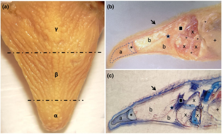
FIGURE 2.

Gross photograph showing (a) macroscopical aspect of the blackspot seabream tongue, which is divided in three areas: the apex (α), the body (β) and the root (γ). Stereomicrographs (b and c) sagittal sections of the tongue: The arrow indicates the tongue dorsal surface covered by several papillae. At the apex level, a pad of mesenchimal tissue is evident (a), with aborally a cartilagineous tissue (asterisk). From the aboral portion of cartilagineous tissue, bone trabeculae were evident, originated by indirect ossification, delimitating niches of adipose tissue (b). An area of compact bone tissue (star) originates through a process of indirect ossification to replace a pre‐existing cartilagineous tissue (square). Adipose tissue (×), areas of hyaline cartilage (circles) and gills (+) characterize the aboral areas of the tongue stratigraphy
