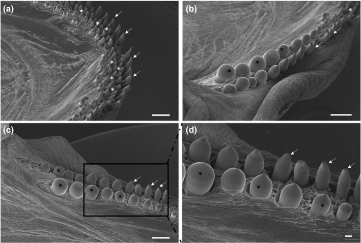
FIGURE 3.

(a) Scanning electron micrograph of the rostral part of the oral cavity with cardiform (white asterisks) and caniniform teeth with aborally curved tips (arrows). (b and c) The molariphorm (black asterisks), caniniform (arrows) and cardiform (white asterisk) teeth on the oral cavity margins. (d) At higher magnification, the molariform (asterisks) and caniniform (arrows) teeth. Scale bar: a, b, c: 1mm; d: 200 μm
