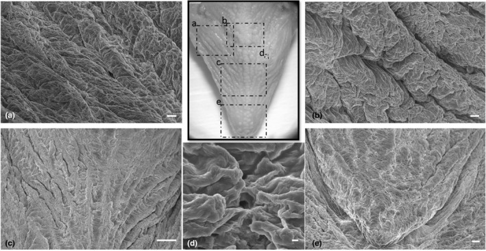
FIGURE 4.

(a–c,). Scanning electron micrograph of the irregular dorsal surface of the tongue with prominent folds and deep furrows, with a latero‐medial orientation fading towards the apex. (d) Taste pores were observed among the folds. (e) The dorsal surface of the apex with less prominent folds. Scale bar: a, b: 100 μm; c: 1mm; d: 10 μm; e: 200 μm
