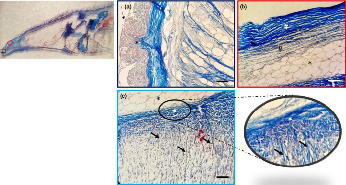
FIGURE 5.

Light micrographs (Masson's trichrome with aniline blue staining): (a) In the apex, a weakly keratinized stratified squamous epithelium (arrow) and connective papillae (asterisk), showing between the epithelial laminae. (b) The dense fibrillar connective tissue of the papillae with abundant collagen fibres (white hashtag) and the deeper loose fibrillar connective tissue (black hashtag), which continued with a pad of unilocular adipose tissue (asterisk). (c) Unilocular adipose tissue (black asterisk); the dense connective tissue (white asterisk) forms septa (arrows) outlining niches of cells, at higher magnification in the insert. Scale bar: 20 μm
