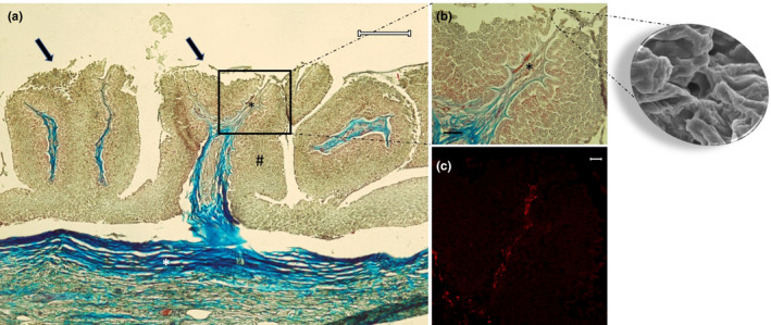
FIGURE 7.

Light micrographs (Masson's trichrome with aniline blue staining) showing (a) in the body and root fungiform‐like papillae (arrows). The mucosa (hashtag) raises in plicae showing the papillae. The connective tissue (white asterisk) throws itself in the mucosa bringing vessels and nerves (black asterisk). (b) A taste pore showed also in the scanning electron micrograph insert, and vessels and nerve (asterisk). (c) Note the presence of papillae with S100 positive nervous fibres. Scale bar: a: 200 μm; b: 30 μm; c: 20 μm
