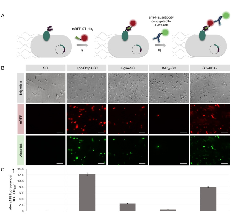
Figure 3.
Investigation on functionality and surface exposure of SC fused to different membrane anchors. E. coli cells expressing the SC intracellularly or as genetic fusion to the membrane anchors Lpp‐OmpA, PgsA, INPNC or AIDA‐I were analyzed 20 h after induction of protein expression. (A) Cells were incubated with a purified mRFP‐ST‐his6 protein that covalently binds to functional SC and surface localization was confirmed using a membrane‐impermeable Alexa488 conjugated anti‐his6 antibody. The stained cells were analyzed using fluorescence microscopy (B) and the Alexa488 fluorescence of the different cell suspensions was quantified using a microplate reader in order to compare the display capacities (C). Scale bars correspond to 10 μm. Error bars were obtained from at least two independent experiments. Please note that the aberrant shape of cells expressing SC intracellularly could be due to the use of the pET‐EXPn1 vector as discussed in more detail in Figure S5.

