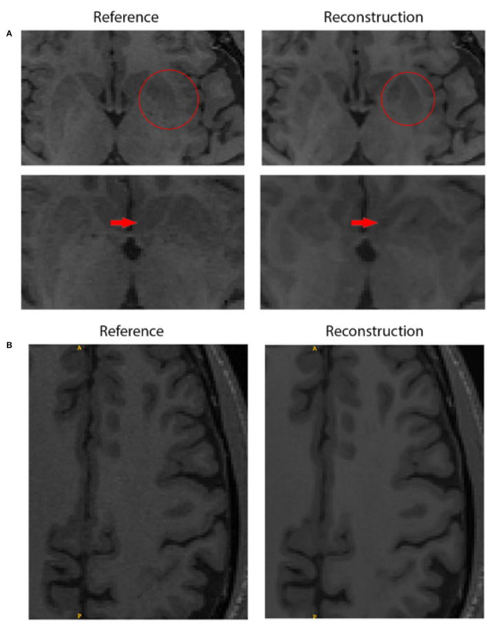
Figure 2.
Quality assessment comparing the fully sampled reference and the reconstruction obtained by team ResoNNance 2.0. (A) The top row shows the border of the left putamen, where the reconstructed image has a discrepancy in shape compared to the reference image (highlighted with red circles). The bottom row shows that changes in the shape of the structure are also visible in the next slice of the same subject (highlighted with red arrows). It is important to emphasize that these discrepancies are not restricted to the putamen, but a systematic evaluation of where these changes occur is out of scope for this work. (B) Illustration of a case where the expert observed rated that the deep-learning-based reconstruction improved image quality. In this figure, we can see smoothening of cortical white matter without loss of information as no changes appeared in the pattern of gyrification within cortical gray matter.

