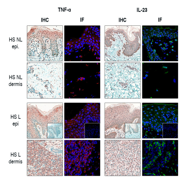
Figure 3.

Immunofluorescent staining further confirms the epidermal presence of IL‐23 and TNF‐α in HS. The presence of IL‐23 and TNF‐α in non‐lesional HS samples was validated by the parallel application of IHC and IF staining in HS samples. IL‐23 and TNF‐α staining was prominent in the epidermis of non‐lesional HS, while their dermal occurrence was weak. In lesional HS, IL‐23+ and TNF‐α+ dermal cell counts were remarkable, while epidermal presence became even more pronounced. Negative control staining is presented in the bottom right corner of images in the third row.
