
Figure 4.
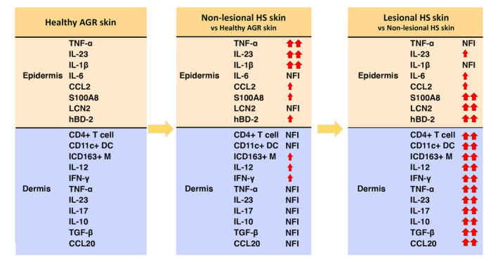
Keratinocytes can be considered as the key driver cells in HS pathophysiology According to our findings, we propose that epidermal, KC‐mediated immune activity is the first step in HS development since all investigated AMPs and the pro‐inflammatory cytokines, IL‐1β, IL‐23 and TNF‐α, are already highly expressed in non‐lesional HS skin by KCs. During the disease progression, when HS lesions develop, the dermal production of IL‐23 and TNF‐α is also significantly enhanced supplemented with an increased influx of inflammatory cells and elevated protein levels of Th1/Th17‐related cytokines and chemokines in the dermis. At the same time, the epidermal presence of IL‐1β, IL‐23 and TNF‐α proteins remains high without further significant increase, compared with non‐lesional HS. Altogether, these results confirm KCs as the key driver cells of HS pathogenesis. The data presented are based on our findings at the protein level. Small red arrows mean nonsignificant upregulation with FC ≥ 2, while duplicated bigger red arrows indicate significant upregulation. NFI means ‘no further increase’ and represents nonsignificant changes with fold change (FC) lower than 2. Abbreviations: AGR, apocrine gland‐rich; CD163+ M, CD163+ macrophage; DC, dendritic cell; HS, Hidradenitis suppurativa; HS‐L, lesional HS; HS‐NL, non‐lesional HS; IF, immunofluorescence; IHC, immunohistochemistry; KC, keratinocyte).