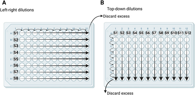
Figure 3.

Two configurations for serial dilutions of serum and mAb samples. Left‐right dilutions (A) are best for samples with expected higher antibody titers. Top‐down dilutions (B) are best for processing more samples with lower expected antibody titers. S1‐S12 denote samples 1‐12. Regardless of configuration, serum samples are diluted two‐fold by transferring 120 μl to 120 μl infection medium, and mAb samples are diluted three‐fold by transferring 80 μl to 160 μl infection medium. An equal volume (120 or 80 μl) is removed from the last column or row.
