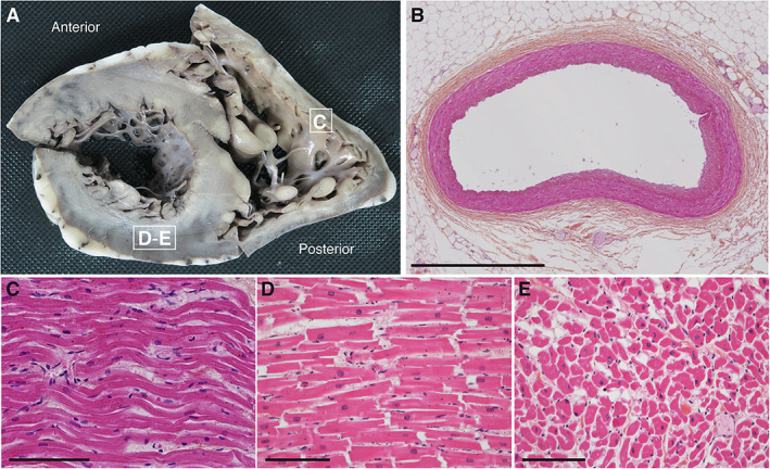
FIGURE 1.

Macroscopic and microscopic pictures of the heart autopsy of Patient 1. (A) Short axis view of a transverse section of the right and left ventricles. Morphological cardiovascular examination including valves and coronary arteries inspection revealed no gross abnormalities. White squares indicate sites of sections used for histological analysis in C–E. (B–E) Hematoxylin and eosin–stained transversal sections of the anterior interventricular artery (B), longitudinal sections of the right ventricle myocardium (C), the posterior wall of the left ventricle myocardium (D), and cross‐sectional section of the left ventricle (E). No contraction band necrosis was detected. Scale bars represent 1mm (B) or 100μm (C–E).
